

A cross sectional study to assess the prevalence of pubertal signs among primary school girls aged 6–8 years in Najaf Governorate, Iraq
دراسة مقطعية لتقييم انتشار علامات البلوغ لدى تلميذات المدارس الابتدائية بعمر 6–8 سنوات في محافظة النجف، العراق
Sajad A Algazali1
1 Department of Pathological Analysis, College of Science, University of Kufa, Najaf, Iraq
Email: sajad.alwan.kitap@uomus.edu.iq
DOI: https://doi.org/10.53796/hnsj76/79
Arabic Scientific Research Identifier: https://arsri.org/10000/76/79
Volume (7) Issue (6). Pages: 1428 - 1436
Received at: 2026-05-15 | Accepted at: 2026-05-22 | Published at: 2026-06-01
Abstract: Background: It has been reported around the world that girls reach puberty at an early age, but there is scant school-based information about early puberal development in Iraq, particularly for girls. Objectives: This study aimed at estimating the prevalence and pattern of the pubertal signs in primary school girls aged 6-8 years in Najaf Governorate. Design: This is a cross sectional study with 351 girls from Najaf recorded and 341 girls analysed, the 9 girls with implausible height and 1 missing age were excluded. The following measurements were recorded: breast development, presence of pubic hair, and presence of axillary hair were recorded as present or absent. All prevalence values were given as Wilson 95% confidence intervals and comparisons were made between groups using chi-square, Fisher's exact and Mann–Whitney tests. Results: At least one pubertal sign was present in 7/341 (2.1%; 95% CI 1.0–4.2) of girls and breast development in 3/341 (0.9%; 95% CI 0.3–2.6). No girl was age 6 years when she got a sign. Signs were found in 4/142 (2.8%; 95% CI 1.1–7.0) of girls aged 7 years and 3/119 (2.5%; 95% CI 0.9–7.2) of girls aged 8 years (trend p = 0.27). For girls less than 8 years old, there were no girls with breast development (0/222; upper 95% limit 1.7%) and four girls with pubertal signs had only pubic hair present. The three girls that were developing breast were 8 years of age. Girls with signs had a higher BMI than girls without signs (21.5 ± 4.0 versus 17.9 ± 3.4 kg/m²; p = 0.014). Conclusion: Pubertal signs were rare in girls in the age of 6-8 years and before 8 years was no breast development observed in Najaf. Due to small numbers of affected girls and the less than optimal quality of measurement, the estimates should be validated in a larger study using standardized examination and exact dates of birth.
Keywords: precocious puberty; early puberty; thelarche; adrenarche; schoolgirls; Iraq.
المستخلص: الخلفية: تشير تقارير من أنحاء العالم إلى بدء البلوغ لدى الفتيات في سن مبكرة، إلا أن البيانات المدرسية عن التطور البلوغي المبكر في العراق لا تزال محدودة. الأهداف: تقدير انتشار علامات البلوغ وأنماطها لدى تلميذات المدارس الابتدائية بعمر 6–8 سنوات في محافظة النجف. المنهجية: دراسة مقطعية سُجّلت فيها بيانات 351 تلميذة، وحُلّلت بيانات 341 منهن بعد استبعاد تسع حالات ذات أطوال غير معقولة وحالة تفتقر إلى العمر. سُجّل وجود أو غياب نمو الثدي وشعر العانة وشعر الإبط. حُسبت فواصل الثقة بنسبة 95% بطريقة ويلسون، واستُخدمت اختبارات كاي تربيع وفيشر الدقيق ومان–ويتني للمقارنات. النتائج: وُجدت علامة بلوغ واحدة على الأقل لدى 7/341 تلميذة (2.1%؛ فاصل الثقة 95%: 1.0–4.2)، ونمو الثدي لدى 3/341 (0.9%؛ 0.3–2.6). لم تُرصد علامات لدى ذوات الست سنوات، مقابل 4/142 بعمر سبع سنوات (2.8%؛ 1.1–7.0)، و3/119 بعمر ثماني سنوات (2.5%؛ 0.9–7.2)، وكانت قيمة احتمال الاتجاه 0.27. لم يُرصد نمو الثدي لدى من هن دون الثامنة (0/222؛ الحد الأعلى لفاصل الثقة 95%: 1.7%)، واقتصرت العلامات لدى أربع منهن على شعر العانة. وكانت الحالات الثلاث لنمو الثدي بعمر ثماني سنوات. ارتفع مؤشر كتلة الجسم لدى ذوات العلامات مقارنة بغيرهن (21.5 ± 4.0 مقابل 17.9 ± 3.4 كغ/م²؛ p = 0.014). الاستنتاج: كانت علامات البلوغ نادرة بعمر 6–8 سنوات، ولم يُلاحظ نمو الثدي قبل الثامنة في عينة النجف. ونظرًا لقلة الحالات ومحدودية جودة القياسات، ينبغي التحقق من التقديرات بدراسة أكبر تعتمد فحصًا موحدًا وتواريخ ميلاد دقيقة.
الكلمات المفتاحية: البلوغ المبكر؛ البلوغ في سن مبكرة؛ بدء نمو الثدي؛ بدء نشاط الغدة الكظرية؛ تلميذات المدارس؛ العراق.
Introduction
In many countries the age at which girls begin puberty has decreased over the last few decades. Meta-analyses including studies from multiple regions have shown significant decreases in the age at breast development (which is an indicator of the timing of puberty) from 1977 to 2013 [1] and cohort studies in Denmark and the United States have shown a similar pattern [2–4]. It also has clinical and public-health implications, with earlier puberty being linked with psychosocial problems and early menarche and decreased adult height with a very precocious onset [5].
Puberty is considered precocious for girls when their secondary sex characteristics appear before age 8 years. [5,6]. Most girls with true central CP have no identifiable cause [7] while the presence of axillary or pubic hair without breast development is typically considered to be due to premature adrenarche, not activation of the hypothalamic–pituitary–gonadal axis [7,8]. Each of these signs of puberty needs to be recorded separately and the stages used as described by Marshall and Tanner [9] to differentiate these patterns. There have been many reports of increased BMI being associated with earlier breast development [10,11].
The figures of diagnosed precocious puberty based on registers in Denmark are of the order of 0.2% in girls [12] but these do not reflect the clinical signs that are seen by families and school doctors. There are no published school-based estimates of pubertal signs in young girls from central and southern Iraq, so there is less evidence to guide clinical pathways and public-health planning.
We thus aimed at (i) estimating the prevalence of pubertal signs in the age group of 6-8 years of girls enrolled in primary schools in Najaf Governorate, (ii) describing the pattern of individual signs and (iii) comparing the anthropometric (growth and development) characteristics of girls with and without signs.
Methods
Study design and setting
This was a cross sectional study of primary school girls (1st to 3rd grades) in the city of Najaf in Iraq, a school-based study. The sampling method used in selection of the primary schools of the study was multistage cluster sampling and convenience sampling method with data collection period from 10 October/2025 to 10 January/2026, involving 351 (one hundred and fifty-one) primary schools. This report follows the recommendations for cross sectional studies, STROBE.
Participants
The age range was 6-8 years old for girls. 351 girls were recorded in Najaf. In addition, another 80 girls were recorded in Babylon Governorate but were not included in the current analysis due to the small number in this sub-sample and the inability to demonstrate that it could be compared with the assessment of the Najaf sub-sample. Prior to analysis, 10 records from Ten Najaf were excluded, all of which were records of a girl shorter than 85 cm: these dates tended to bunch together in a small number of contiguous records, and 1 was missing from an age date. This final sample consisted of 341 girls. No exclusions of girls with a pubertal sign were made, so there was no impact of the exclusions on the prevalence estimates (see Sensitivity analyses).
The collection and definitions of data are as follows:
Parental/guardian or school staff provided ages, grades, chronic illness, regular medications and current treatment that impacts pubertal development. Weight (kg) and height (cm) were both measured and BMI was calculated by dividing weight (kg) by the height (cm)2. Pubertal signs were assessed by the resident (a physician with a special interest in infertility and physiology) and breast development, pubic hair and axillary hair were scored as present (Tanner stage 2 or higher) or absent.
The main outcome, “pubertal signs”, was considered to be the presence of at least one of the following stages: breast development (thelarche), pubic hair (pubarche) or axillary hair. Since the earliest sign of a tonically active gonadal axis in most girls is the development of their breast, the development of the breast was analysed separately, while the stage of pubic and axillary hair development was regarded as an adrenarche-type pattern. The signs were described as being “suggestive of precocious development” for girls below 8 years, not a diagnosis. One girl had no record of any of the three signs, only a general “pubertal signs”, and was deemed negative in the primary analysis and positive in a sensitivity analysis.
Statistical analysis
All values are expressed as mean ± SD or number (%). Prevalence is given with Wilson 95% confidence intervals. The chi-square test, Cochran–Armitage trend test and Fisher’s exact test were used to compare proportions across age groups, while the Mann–Whitney U test was used to compare continuous variables between groups of girls with and without signs. As there were few girls with signs (n = 7), no multivariable regression was performed. All tests were conducted at the 5% level. Python 3 with SciPy and statsmodels software was used for analysis.
Results
Participants
Of 351 Najaf girls recorded, 341 were analysed (Figure 1): 80 aged 6 years, 142 aged 7 years and 119 aged 8 years (mean age 7.1 ± 0.8 years) (Table 1). A single girl (0.3%) was identified with a chronic condition (diabetes) and a single girl was identified as using regular medicines (asthma) while a single girl was identified as obese and a single girl was identified as having anemia. One girl who had all 3 pubertal signs was already being treated.
Table 1. Characteristics of the girls included in the analysis, overall and by age
| Characteristic | 6 years (n = 80) | 7 years (n = 142) | 8 years (n = 119) | All (n = 341) |
|---|---|---|---|---|
| First grade, n (%) | 77 (96.3) | 51 (35.9) | 3 (2.5) | 131 (38.4) |
| Second grade, n (%) | 3 (3.8) | 91 (64.1) | 66 (55.5) | 160 (46.9) |
| Third grade, n (%) | 0 (0.0) | 0 (0.0) | 50 (42.0) | 50 (14.7) |
| Weight, kg | 19.7 ± 2.7 | 21.7 ± 3.9 | 26.9 ± 5.1 | 23.0 ± 5.0 |
| Height, cm | 111.1 ± 11.5 | 109.5 ± 11.8 | 120.1 ± 9.7 | 113.6 ± 12.0 |
| BMI, kg/m² | 16.3 ± 3.3 | 18.3 ± 3.5 | 18.7 ± 3.1 | 18.0 ± 3.4 |
| Chronic illness, n (%) | 0 (0.0) | 1 (0.7) | 3 (2.5) | 4 (1.2) |
| Regular medication, n (%) | 0 (0.0) | 0 (0.0) | 2 (1.7) | 2 (0.6) |
| ≥1 pubertal sign, n (%) | 0 (0.0) | 4 (2.8) | 3 (2.5) | 7 (2.1) |
Values are mean ± SD unless stated. BMI, body mass index. Height and BMI are based on 340 girls (height missing for one).
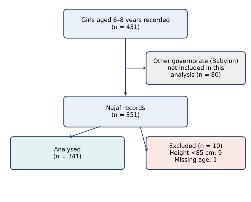 Figure 1. Flow of participants from recording to analysis.
Prevalence of pubertal signs
At least one pubertal sign was present in 7/341 (2.1%; 95% CI 1.0–4.2) of girls. Breast development was found in 3/341 (0.9%; 95% CI 0.3–2.6), pubic hair in 7/341 (2.1%; 95% CI 1.0–4.2) and axillary hair in 2/341 (0.6%; 95% CI 0.2–2.1). Of the seven girls who showed signs, 4 had only pubic hair, 1 had only breast development and pubic hair, and 2 had all three signs (Table 4, Figure 3).
No girl aged 6 years had a sign (0/80). There was no statistical difference in prevalence between 7 and 8 years (chi-square, p = 0.33; trend, p = 0.27; Fisher’s exact, 8 years versus younger, p = 0.70) (Table 2, Figure 2). Among girls younger than 8 years (n = 222), 4/222 (1.8%; 95% CI 0.7–4.5) had at least one sign, all four with isolated pubic hair, and none had breast development (0/222; 95% CI 0.0–1.7). The three girls who had developed breast were 8 years old.
Table 2. Prevalence of pubertal signs by age
| Sign, n (%) | 6 y (n = 80) | 7 y (n = 142) | 8 y (n = 119) | <8 y (n = 222) | All (n = 341) |
|---|---|---|---|---|---|
| ≥1 sign | 0 (0.0) | 4 (2.8) | 3 (2.5) | 4 (1.8) | 7 (2.1) |
| Breast development | 0 (0.0) | 0 (0.0) | 3 (2.5) | 0 (0.0) | 3 (0.9) |
| Pubic hair | 0 (0.0) | 4 (2.8) | 3 (2.5) | 4 (1.8) | 7 (2.1) |
| Axillary hair | 0 (0.0) | 0 (0.0) | 2 (1.7) | 0 (0.0) | 2 (0.6) |
95% confidence intervals for the overall prevalence: ≥1 sign 1–4.2%; breast development 0.3–2.6%. y, years.
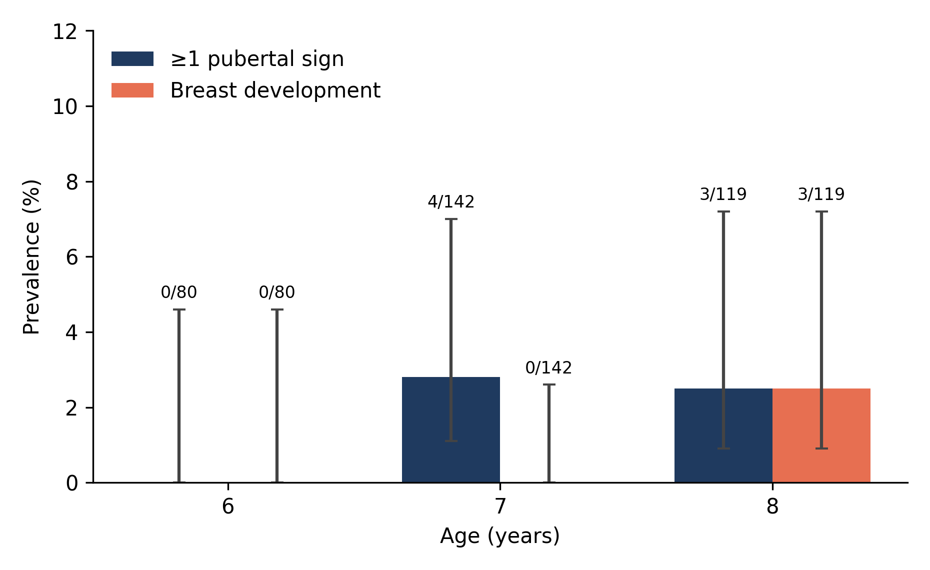
Figure 2. Prevalence of at least one pubertal sign and of breast development by age. Bars show the percentage with Wilson 95% confidence intervals; numbers above bars are cases/total.
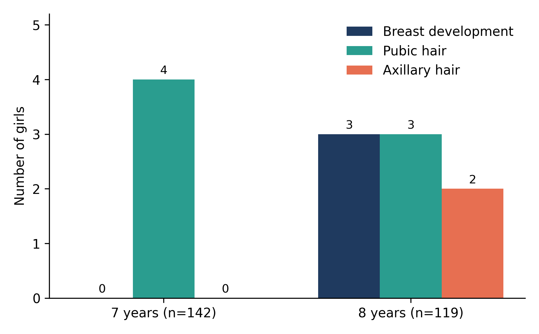
Figure 3. Number of girls with each pubertal sign at ages 7 and 8 years (none at age 6).
Anthropometric comparison
Girls with pubertal signs were slightly older than girls without signs (7.4 ± 0.5 versus 7.1 ± 0.8 years) and had a higher BMI (21.5 ± 4.0 versus 17.9 ± 3.4 kg/m²; p = 0.014) (Table 3, Figure 4). Weight and height did not differ significantly. Given that only seven girls had signs, these comparisons are imprecise and were not adjusted for age.
Table 3. Comparison of girls with and without pubertal signs
| Variable | ≥1 sign (n = 7) | No signs (n = 334) | p-value* |
|---|---|---|---|
| Age, years | 7.4 ± 0.5 | 7.1 ± 0.8 | 0.291 |
| Weight, kg | 26.4 ± 11.0 | 23.0 ± 4.9 | 0.827 |
| Height, cm | 109.3 ± 21.1 | 113.7 ± 11.8 | 0.272 |
| BMI, kg/m² | 21.5 ± 4.0 | 17.9 ± 3.4 | 0.014 |
Values are mean ± SD. *Mann–Whitney U test.
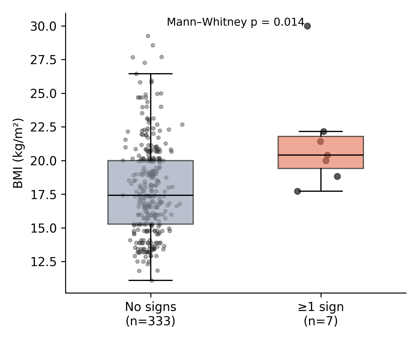
Figure 4. Body mass index in girls with and without pubertal signs. Boxes show the median and interquartile range; points are individual girls.
Table 4. Pattern of pubertal signs in the seven girls with at least one sign
| Girl | Age, y | Grade | BMI, kg/m² | Breast | Pubic hair | Axillary hair |
|---|---|---|---|---|---|---|
| 1 | 7 | 2 | 20.0 | No | Yes | No |
| 2 | 7 | 2 | 18.8 | No | Yes | No |
| 3 | 7 | 2 | 17.7 | No | Yes | No |
| 4 | 7 | 2 | 22.2 | No | Yes | No |
| 5 | 8 | 2 | 30.0 | Yes | Yes | Yes |
| 6 | 8 | 3 | 20.4 | Yes | Yes | Yes |
| 7 | 8 | 3 | 21.4 | Yes | Yes | No |
y, years. Girls 1–4 were recorded consecutively from the same second-grade class (see Discussion).
Sensitivity analyses
Retaining all Najaf records with known age gave 7/350 (2.0%; 95% CI 1.0–4.1). If the girl was excluded from the analysis because of a non-specific general “pubertal signs” entry, the result was 6/340 (1.8%; 95% CI 0.8–3.8), which is considered negative; if the girl with a general “pubertal signs” entry was counted as positive because there was no specific entry, the result was 8/341 (2.3%; 95% CI 1.2–4.6), which is considered positive. Restricting the analysis to girls with a recorded height of at least 100 cm gave 4/291 (1.4%; 95% CI 0.5–3.5). Excluding the four girls who had only pubic hair and were recorded consecutively at 7 years, affected 3/337 (0.9%; 95% CI 0.3–2.6), and no girl under 8 years old was affected (0/218).
Discussion
Of the 341 girls aged 6-8 years in this school based sample from Najaf, 2.1% had at least one pubertal sign and 0.9% had breast development. None of the girls aged 6 had a sign, all three girls who had developed breast development were of age 8 years. Therefore, not many pubertal signs were present and no signs of genuine gonadal activation prior to the standard age limit of 8 years [5,6] were noted.
Of the 222 girls younger than 8 years, 4 of them had pubic hair only and none of the girls had breast development (upper 95% confidence limit 1.7%). Isolated pubic hair without thelarche is the most typical presentation of ‘premature adrenarche’ or ‘pubarche’, which is generally not serious and should be clinically evaluated [7,8]. We have also reported signs separately in order to avoid pooling this distinction. Our figures cannot be compared directly to the register data on diagnosed precocious puberty [12] as the former are screening observations without confirmation, whereas the latter are the data of referred and diagnosed girls.
There was no significant difference between the prevalence for girls 7 and 8 years of age. This is unlike the steep age gradient observed in other populations [1-4] and likely related to the small number of events and the recording pattern described below, along with the use of completed years of age. In this population, a larger sample size, standardization of age at onset by the examiner and birth dates would be required to describe the age at onset.
BMI was also greater in girls with signs and this finding is consistent with the reported association between adiposity and breast development at an earlier age [10,11]. This finding is based on seven girls and has not been age-adjusted and is based on heights that were not always reliable (see below). Thus, we regard it as hypothesis-generating. BMI-for-age z-scores [13] were not computed, and thus age and sex-specific norms were not taken into consideration.
There are some data quality problems that should be highlighted. First, four of the seven girls with signs were recorded consecutively from the same second-grade class, were aged 7 years, had isolated pubic hair and had recorded heights of 95–100 cm, values that are very low for their age. This pattern can be due to an actual cluster, artefact in recording or measurement or inaccurate age. The prevalence of 0.9% following exclusion is unlikely to be affected significantly by the exclusion of girls who had breast development before 8 years, and the estimate of isolated pubic hair at 7 years is somewhat fragile. Second, there was a recorded 49 girls (14%) with a height below 100 cm, and 62% of the heights were multiples of 5 cm, implying rounded or estimated heights, and limiting anthropometric inference.
Clinically, the results indicate that girls with such early signs of puberty should be referred for clinical evaluation and, if necessary, for hormonal and bone age evaluation, but that these early signs are rare in this age group from the public health standpoint in Najaf. The study also demonstrates the necessity of standardizing examination methods in order to be able to follow up on pubertal signs in Iraq in the future.
Strengths and limitations
Strengths are that it contains a school-based sample of over three hundred girls, breast, pubic and axillary sites were recorded separately, and the implausible data were handled transparently, with sensitivity analyses indicating the stability of the main estimate. Acknowledging limitations is a good thing to do. First, only seven girls were found to have signs, which limits the precision of prevalence estimates (95% CI 1.0-4.2%) and multivariable analysis was not possible. Second, the design was cross-sectional, rather than longitudinal, meaning that it did not measure onset of signs, but rather age at onset; Ages at onset were recorded in completed years, which means that exact classification around the 8-year onset is not possible. Third, there were no hormonal or bone-age confirmations available, so the presence of signs in girls younger than 8 years old implies suspected, but not confirmed, precocious development. Fourth, some findings may be confounded by the potential for clustering from one class, and by the quality of the height measurements. Fifthly, family history, maternal factors and diet and environmental exposures (endocrine disrupting chemicals) were not analysed. Sixth, the sample is from one governorate and cannot be assumed to be representative of Iraq.
Conclusion
Breast development before age 8 was not observed (0/222) and puberal signs were uncommon (2.1%) among girls aged 6–8 years in Najaf. The signs at age 7 years were isolated pubic hairs, an adrenarche-type hair pattern and are subject to confirmation due to the possible clustering and measurement error. More uniform examination, dates of birth and BMI z-scores, and hormonal confirmation of suspected cases in a larger multicentre study, are recommended.
Data availability: Data underlying this article are available on reasonable request from the corresponding author.
References
1. Eckert-Lind C, Busch AS, Petersen JH, Biro FM, Butler G, Bräuner EV, et al. Worldwide secular trends in age at pubertal onset assessed by breast development among girls: a systematic review and meta-analysis. JAMA Pediatr. 2020;174(4):e195881.
2. Aksglaede L, Sørensen K, Petersen JH, Skakkebæk NE, Juul A. Recent decline in age at breast development: the Copenhagen Puberty Study. Pediatrics. 2009;123(5):e932–9.
3. Herman-Giddens ME, Slora EJ, Wasserman RC, Bourdony CJ, Bhapkar MV, Koch GG, et al. Secondary sexual characteristics and menses in young girls seen in office practice: a study from the Pediatric Research in Office Settings network. Pediatrics. 1997;99(4):505–12.
4. Biro FM, Greenspan LC, Galvez MP, Pinney SM, Teitelbaum S, Windham GC, et al. Onset of breast development in a longitudinal cohort. Pediatrics. 2013;132(6):1019–27.
5. Carel JC, Léger J. Precocious puberty. N Engl J Med. 2008;358(22):2366–77.
6. Kaplowitz PB, Oberfield SE. Reexamination of the age limit for defining when puberty is precocious in girls in the United States: implications for evaluation and treatment. Pediatrics. 1999;104(4):936–41.
7. Latronico AC, Brito VN, Carel JC. Causes, diagnosis, and treatment of central precocious puberty. Lancet Diabetes Endocrinol. 2016;4(3):265–74.
8. Abreu AP, Kaiser UB. Pubertal development and regulation. Lancet Diabetes Endocrinol. 2016;4(3):254–64.
9. Marshall WA, Tanner JM. Variations in pattern of pubertal changes in girls. Arch Dis Child. 1969;44(235):291–303.
10. Kaplowitz PB, Slora EJ, Wasserman RC, Pedlow SE, Herman-Giddens ME. Earlier onset of puberty in girls: relation to increased body mass index and race. Pediatrics. 2001;108(2):347–53.
11. Lee JM, Appugliese D, Kaciroti N, Corwyn RF, Bradley RH, Lumeng JC. Weight status in young girls and the onset of puberty. Pediatrics. 2007;119(3):e624–30.
12. Teilmann G, Pedersen CB, Jensen TK, Skakkebæk NE, Juul A. Prevalence and incidence of precocious pubertal development in Denmark: an epidemiologic study based on national registries. Pediatrics. 2005;116(6):1323–8.
13. de Onis M, Onyango AW, Borghi E, Siyam A, Nishida C, Siekmann J. Development of a WHO growth reference for school-aged children and adolescents. Bull World Health Organ. 2007;85(9):660–7.
 English
 Arabic