
In Vitro Cytotoxicity Potential of Curcumin Nanoparticles Against Human Cancer Cell Line
الإمكانات السمية الخلوية لجسيمات الكركمين النانوية ضد خط خلايا السرطان البشري
Ibtihal Riyadh Najeeb1
1 Department of pathological analyses,Collage of science,University of Kufa,Iraq.
*Corresponding Author E-mail: ibtihalr.alrammahi@uokufa.edu.iq
DOI: https://doi.org/10.53796/hnsj75/70
Arabic Scientific Research Identifier: https://arsri.org/10000/75/70
Volume (7) Issue (5). Pages: 1249 - 1257
Received at: 2026-04-15 | Accepted at: 2026-04-22 | Published at: 2026-05-01
Abstract: Breast cancer remains a major global health challenge, highlighting the need for safer and more effective therapeutic approaches. Curcumin has attracted considerable scientific interest because of its antioxidant, anti-inflammatory, and anticancer properties; however, its clinical application is limited by poor aqueous solubility, low bioavailability, and limited cellular uptake. This study aimed to synthesize curcumin nanoparticles using a modified sol–oil method, characterize their physicochemical properties, and evaluate their in vitro cytotoxic activity against the human breast cancer cell line MCF-7. The prepared nanocurcumin was characterized using morphological and structural techniques, including atomic force microscopy, field emission scanning electron microscopy, X-ray diffraction, Fourier-transform infrared spectroscopy, and UV-visible spectroscopy. The results confirmed the successful formation of stable curcumin nanoparticles with nanoscale dimensions, quasi-spherical morphology, improved dispersibility, and preserved chemical structure. The anticancer activity was assessed using the MTT assay after exposure of MCF-7 cells to different concentrations of nanocurcumin for 72 hours. The findings revealed a clear concentration-dependent cytotoxic effect, with inhibition rates increasing from 35.47% at 6.25 µg/mL to 79.66% at 100 µg/mL. The calculated IC50 value was approximately 14 µg/mL, indicating strong antiproliferative activity against breast cancer cells. These results suggest that nanoscale formulation enhances the biological effectiveness of curcumin, likely by improving solubility, cellular uptake, and interaction with tumor cells. The study concludes that curcumin nanoparticles possess promising physicochemical and anticancer properties and may represent a potential candidate for breast cancer therapy. Further molecular, in vivo, and safety studies are recommended to clarify their mechanism of action and evaluate their suitability for future clinical applications.
Keywords: Curcumin nanoparticles; Nanocurcumin; MCF-7 cell line; Breast cancer; Cytotoxicity assay.
المستخلص: لا يزال سرطان الثدي يمثل تحديًا صحيًا عالميًا كبيرًا، مما يبرز الحاجة إلى تطوير استراتيجيات علاجية أكثر أمانًا وفعالية. وقد حظي الكركمين باهتمام علمي واسع نظرًا لخصائصه المضادة للأكسدة والالتهاب والسرطان؛ إلا أن استخدامه السريري يظل محدودًا بسبب ضعف ذوبانيته في الماء، وانخفاض توافره الحيوي، ومحدودية امتصاصه الخلوي. هدفت هذه الدراسة إلى تحضير جسيمات الكركمين النانوية باستخدام طريقة الزيت–السول المعدلة، وتوصيف خصائصها الفيزيائية والكيميائية، وتقييم فعاليتها السمية الخلوية مختبريًا ضد خط خلايا سرطان الثدي البشري MCF-7. جرى توصيف النانوكركمين المحضر باستخدام عدد من التقنيات الشكلية والبنيوية، شملت المجهر القوي الذري، والمجهر الإلكتروني الماسح ذي الانبعاث الحقلي، وحيود الأشعة السينية، ومطيافية الأشعة تحت الحمراء بتحويل فورييه، ومطيافية الأشعة فوق البنفسجية–المرئية. أكدت النتائج نجاح تكوين جسيمات كركمين نانوية مستقرة ذات أبعاد نانوية، وشكل شبه كروي، وقابلية تشتت محسنة، مع الحفاظ على التركيب الكيميائي للكركمين. تم تقييم النشاط المضاد للسرطان باستخدام اختبار MTT بعد تعريض خلايا MCF-7 لتراكيز مختلفة من النانوكركمين لمدة 72 ساعة. أظهرت النتائج تأثيرًا سميًا خلويًا واضحًا يعتمد على التركيز، إذ ارتفعت نسب تثبيط نمو الخلايا من 35.47% عند تركيز 6.25 ميكروغرام/مل إلى 79.66% عند تركيز 100 ميكروغرام/مل. كما بلغت قيمة IC50 المحسوبة نحو 14 ميكروغرام/مل، مما يشير إلى نشاط قوي مضاد لتكاثر خلايا سرطان الثدي. وتشير هذه النتائج إلى أن الصياغة النانوية للكركمين تعزز فعاليته الحيوية، على الأرجح من خلال تحسين الذوبانية، وزيادة الامتصاص الخلوي، وتعزيز التفاعل مع الخلايا الورمية. وتخلص الدراسة إلى أن جسيمات الكركمين النانوية تمتلك خصائص فيزيائية وكيميائية ومضادة للسرطان واعدة، وقد تمثل مرشحًا محتملًا لعلاج سرطان الثدي. وتوصي الدراسة بإجراء مزيد من الأبحاث الجزيئية والدراسات داخل الجسم الحي ودراسات السلامة لتوضيح آلية عملها وتقييم ملاءمتها للتطبيقات السريرية المستقبلية.
الكلمات المفتاحية: جسيمات الكركمين النانوية؛ النانوكركمين؛ خط خلايا MCF-7؛ سرطان الثدي؛ اختبار السمية الخلوية.
-
Introduction
Cancer is still one of the top causes of death in the world. World Health Organization (WHO) estimates state that 8.2 million deaths were caused by cancer in the year 2012, and more than 13 million deaths, occurring until the year of 2030 (zhang et al., 2017). It is likely the result of drug resistance, tumor recurrence, severe side effects and the general therapeutic nonspecificity associated with traditional cancer treatments (i.e. chemotherapy and radiation therapy). Therefore, the emergence of a multitude of breakthrough innovations for safer and more effective treatment infrastructures has been ranked as one of the most critical focal points in cancer research (Liu et al., 2025). Due to the distinct physical and chemical characteristics which nanomaterials possess, recent developments in cancer therapies have investigated the use of nanoparticles as a promising tool offered by nanotechnology during treatment of cancers. Nanoparticles (1 nm–100 nm) increase the solubility, stability, and targeted distribution to tumor tissues, which are a unique advantage of nanocarriers over drug delivery (Ghoran et al., 2022). Nanotechnology can be employed in various fields; however, biomedical and pharmaceutical sciences are two very important fields. Of those, the sol-oil method of generating nanoparticles has gained much interest among various nanosystems since its ease of production process only requires using non-toxic chemicals that may affect future biomedical uses (Gandapu et al., 2011). Curcumin is a naturally occurring polyphenolic compound extracted from the roots of the turmeric plant (Curcuma longa). It has attracted much attention in the literature because of its various biological effects, including antioxidant, anti-inflammatory, antibacterial, and anticancer characteristics (Amaroli et al., 2024). Nanocurcumin can accomplish more pronounced therapeutic properties than natural curcumin owing its high solubility, bioavailability, cellular uptake efficiency with lower doses and greater targeting to affected tissues (Wen et al., 2017). The treatment of small doses of nanocurcumin is more effective on many human cancer cell lines (Basniwal et al., 2014). The present study will synthesize this nanocurcumin using sol-oil method to enhance its solubility, absorption, and bioavailability. Then cytotoxicity and anticancer activity of the compound against human breast cancer cells (MCF-7) will be determined using MTT assay, as a potential conventional therapy alternative.
2. Materials and methods
2.1. Synthesis of curcumin nanoparticles
They are prepared using a modified sol-oil method based on the original study (hanna and Saad, 2020). Curcumin (10 mg) was dissolved in 0.1 mL DMSO and added to 10 mL olive oil under stirring at 600 rpm for 1 h. Next, the solution was sonicated for 2 h at 80 C under ultrasonic conditions, where the sonication amplitude was 5 mm and the pulses were 4 s long with an interval of 1 min between successive pulses. The resulting mixture was directly frozen in liquid nitrogen for 10 min and was then incubated at room temperature for 4 h. The particles formed were obtained by centrifugation at 6000 rpm for 10 min at 4 C, followed by washing with diethyl ether and then dissolved in distilled water and finally freeze-dried to obtain nanocurcumin powder.in nanoparticles (nanocurcumin).
2.2. Characterization of the prepared nanocurcumin
Atomic force microscopy and field emission scanning electron microscopy (FESEM) were used to study the morphological properties of the produced curcumin nanoparticles, as well as X-ray diffraction, infrared spectra, ultraviolet and visible spectra.
2.3. Anticancer activity of curcumin nanoparticles
2.3.1. Maintenance of cell cultures
At the AL-Amin Center for Advanced Biotechnology Research and Techniques in Najaf, the MCF-7 cell line was kept in RPMI-1640 supplemented with 10% fetal bovine, 100 units/mL penicillin, and 100 µg/mL streptomycin. Trypsin-EDTA was used to passage the cells, which were then reseeded twice a week at 80% confluence and cultured at 37 °C (Sulaiman et al., 2018).
2.3.2. Cytotoxicity assays of curcumin nanoparticles
Using 96-well plates, the MTT cell viability assay was used to assess the cytotoxic effect of curcumin nanoparticles. The cells were exposed to the prepared curcumin nanoparticles at different concentrations. After 24 hours and after 72 hours of treatment, the media were removed, 28 microliters of MTT at a concentration of 2 mg/ml were added, and the cells were successfully maintained in the incubator at 37°C. Following the removal of the MTT solution, 130 µL of DMSO (dimethyl sulphoxide) was added to the wells to dissolve the residual crystals. The mixture was then incubated at 37 °C for 15 minutes while being shaken (Jabir et al., 2019). The assay was carried out in triplicate, and the absorbance was measured at a wavelength of 492 nm using an ELISA plate reader. The following formula was used to determine the percentage of cytotoxicity, or the inhibition rate of cell growth:
Cytotoxicity = A-B/A *100……………………….1
Where A the optical density of control and B the optical density of Samples.
3. Results and discussion
3.1. Synthesis of Curcumin nanoparticles
The modified sol-oil approach was successfully used to create curcumin nanoparticles. The combination gradually changed from a clear yellow solution to a turbid yellow suspension during the preparation procedure, signifying the creation of curcumin nanoparticles. A fine yellow precipitate was produced following centrifugation and washing, indicating that nanocurcumin was successfully formed. When compared to raw curcumin, the drying process produced a stable yellow powder that could be redispersed, indicating successful nanoparticle production and enhanced dispersibility.The fast diffusion of curcumin from the organic phase into the oil phase under constant stirring is the primary cause of nanoparticle production in the sol–oil technique. While sonication improves dispersion, decreases particle size, and minimizes aggregation to produce stable nanoscale particles, the use of a hot plate stirrer guarantees uniform mixing.These findings are consistent with earlier research demonstrating that curcumin nanoparticles made using oil-based techniques had superior biological activity and solubility compared to free curcumin. The produced nanocurcumin demonstrated enhanced dispersion and nanoscale particle size, which may improve its cellular uptake and potential for cancer treatment (Hanna and Saad, 2020).
3.2. Morphological characterization of the synthesized Curcumin nanoparticles
Figure 1 shows the atomic force microscopy examination of curcumin nanoparticles. The image above shows the bright white and yellow dots, which represent curcumin nanoparticles distributed on the surface. The XY dimensions indicate that the scan is 10 by 10 meters. The colored bar on the right shows the vertical height of the particles. The highest point recorded in this scan is 3.5 nanometers. This indicates that the particles are very thin or almost flat on the surface, which is a good indicator of size. The lower graph shows the histamine size, where the curved approach shows a very sharp beige peak close to zero, usually between 1 and 4 nanometers, depending on the scale. The narrowness of the curve indicates that the particles are very homogeneous in size, meaning that most of the prepared curcumin particles are approximately the same size and not a mixture of large and small particles. Figure 2 (FESEM) shows the curcumin nanoparticles, revealing a quasi-spherical structure with a uniform distribution. The surface morphology exhibits a coarse texture, likely due to the presence of bioactive organic compounds from the plant extract on the nanoparticle surface. The particle size is 100 nm. The distinct crystalline lattice edges indicate their crystalline nature, as shown in Figure 3. X-ray diffraction (XRD) patterns showed characteristic diffraction peaks at 2θ values of 25–27. The average crystal size, calculated using the Debye–Scherer equation, ranged between 23 and 25 nm, confirming the nanoparticle nature. Figure 4 shows of FTIR, The broad peak (~3400–3500 cm⁻¹): represents the phenolic (O–H) group. In nanoparticles, this peak is often sharpened or slightly altered due to the increased surface area of the molecules, which increases the exposure of hydrogen bonds. The peak at (~1625 cm⁻¹): the most prominent in your image, is due to the overlapping (C=O) and (C=C) double bond vibrations. The fact that this peak remains sharp indicates that the dictone structure of curcumin is unaffected.The peak at (~1510 cm⁻¹): represents the aromatic (C=C) benzene ring vibrations.The peak at (~1270 cm⁻¹): represents the stretching of the (C–O) phenolic bond, a very distinctive marker of curcumin purity.The peak at (~1020–1150 cm⁻¹): represents the methoxy (C–O–C) bond vibrations. Figure 5 shows the ultraviolet and visible spectrum. A clear absorption peak is observed at a wavelength of 420 nm. Nano-curcumin is known for its strong absorption in the blue region of the visible spectrum, between 400-430 nanometers. This explains its orange-yellow color to the human eye. This value confirms that the substance is indeed curcumin and that its chemical structure, specifically the system of alternating double bonds, remains intact after the transformation to the nanoscale. Nano-curcumin often exhibits a slight shift in the apex, with an increase in apex width, compared to regular curcumin.
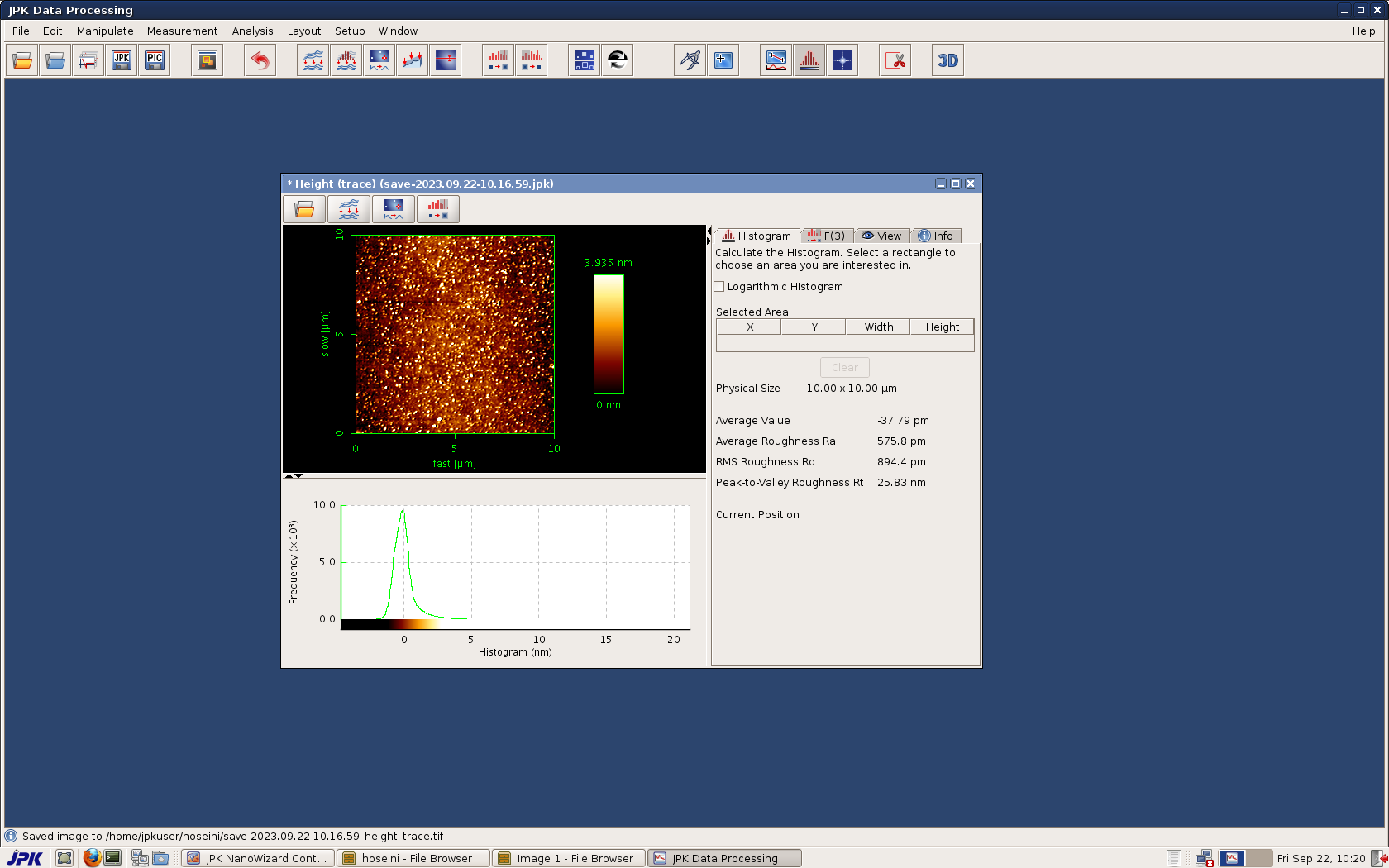
Figure 1: Atomic force microscopy of Curcumin nanoparticles
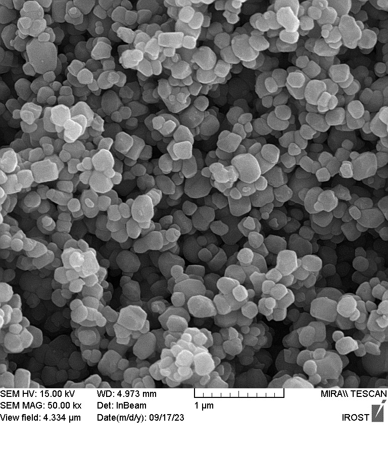
Figure 2 : FESEM of Curcumin nanoparticles
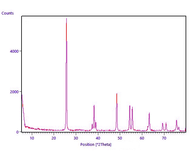
Figure 3: (XRD) spectra of Curcumin nanoparticles
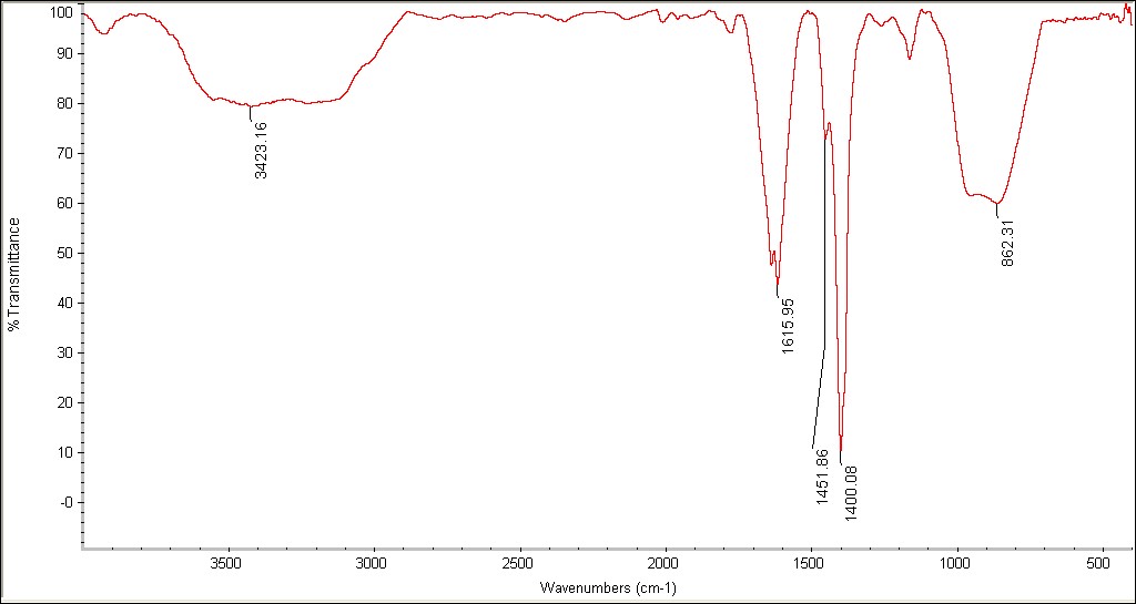
Figure 4: FTIR of Curcumin nanoparticles
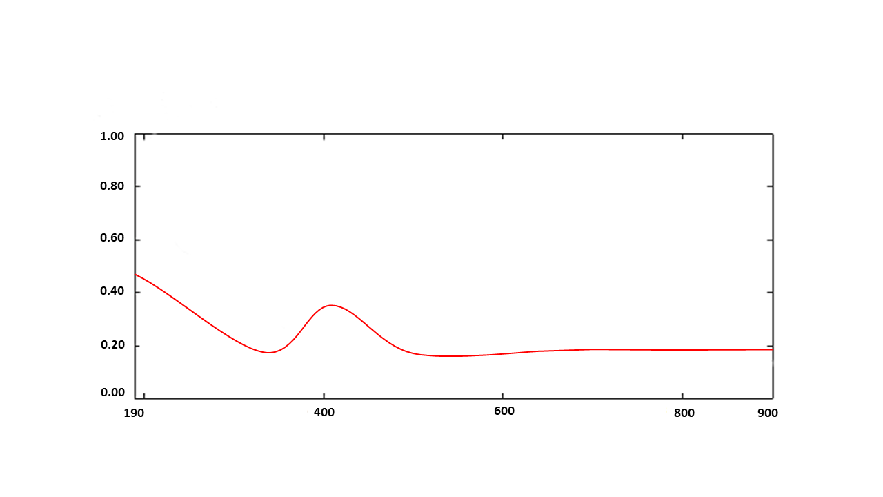
Absorbance a.u.
Wavelength.nm
Figure 5: UV of Curcumin nanoparticles
3.3. Anticancer activity (MTT assay)
3.3.1. Cytotoxicity effect of Curcumin nanoparticles on human of MCF-7 cell
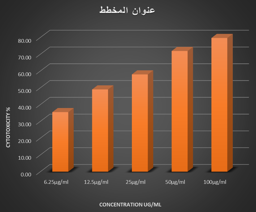
The MTT assay is a colorimetric test that uses cell metabolic activation to cause a color shift. For 72 hours, the cytotoxic effects of curcumin nanoparticles on human cancer cell lines (MCF-7) were investigated. After 72 hours, MCF-7 proliferation was significantly inhibited, according to the data. When compared to the untreated control cells, the cell growth was noticeably reduced. For 72 hours of exposure, the cells were exposed to varying doses of curcumin nanoparticles (6.25, 12.5, 25, 50, and 100 µg/mL). Curcumin nanoparticles exhibited 35.47%, 49%, 57.95%, 71.9%, and 79.66% of the cytotoxicity at 6.25µg/ml, 12.5µg/ml, 25µg/ml, 50µg/ml, and 100µg/ml, respectively. After 72 hours of treatment, curcumin nanoparticles’ estimated IC50 value against MCF-7 cells was roughly 14 µg/mL. (Fig. 6). These results support earlier findings that nanocurcumin has more robust antiproliferative effects than free curcumin in breast cancer models (Elbialy et al., 2013) by confirming that increasing nanoparticle concentrations results in higher inhibition of breast cancer cell growth. The anticancer efficacy of nanocurcumin against MCF-7 cells is strongly supported by studies focused on breast cancer. According to Hosseini et al. (2019), nano-curcumin decreased MCF-7 cell viability by roughly 83.6% at their maximum measured concentration (162.87 mmol/L = 60 mg/mL), with an IC50 value of 59.72 mmol/L ≈ 22 mg/mL. Nanocurcumin showed a similar cytotoxic impact (79% vs. 83.6%) despite the much lower dose utilized in this investigation (100 µg/mL) compared to Hosseini et al. (≈ 60 mg/mL), demonstrating robust antiproliferative action even at significantly lower concentrations. According to Tsai et al. (2025), showed that after 72 hours, polysaccharide-based curcumin nanoparticles showed high cytotoxic effects against MCF-7 cells (IC50 = 17.5 µg/mL), with quick cellular uptake and apoptosis induction. These results are in line with earlier research showing that nanocurcumin has comparable cytotoxic and antiproliferative effects on breast cancer cells. Moreover, similar dose- and time-dependent cytotoxic effects have been seen in different cancer models. According to Hanna and Saad’s (2020) report, the cytotoxicity of nanocurcumin on Hep-2 cells increases with concentration and exposure time, reaching approximately 97.8% cell death at 75µg/mL after 48 hours. The IC50 values decreased from 45.1 µg/ml after 24 hours to 17 µg/ml after 48 hours. This highlights the interplay between exposure time and dose in determining cytotoxic activity. In a similar study, Alam et al. (2022) demonstrated that curcumin-PLGA nanoparticles inhibited proliferation of human gastric cancer cells (AGS cell line) by 97% at a concentration of 40 µM over 72 hours, whereas free curcumin exhibited only 83% inhibition at the same concentration. Greater contact between nanoparticles and biological components is made possible by longer exposure, which disrupts metabolic function and inhibits cell growth.
Our data support the utility of nanocurcumin as a potent anticancer drug with therapeutic flexibility for clinical functioning. Moreover, analogous results have been observed in other studies showing selectivity towards normal cells (Karimi et al., 2024). Numerous in vitro investigations have demonstrated that nanocurcumin is more effective than free curcumin at increasing cytotoxicity and apoptosis in cancer cells, as demonstrated by increased pro-apoptotic gene expression, disruption of cell cycle progression, and accelerated programmed cell death (Hanna & Saad, 2020; Moawad et al., 2023).
 .
.
Figure 6: Cytotoxicity assay of MCF-7 breast cancer cells treated with varying concentrations of curcumin nanoparticles.
4. Conclusions
The sol–oil process was effectively used to create curcumin nanoparticles, producing a stable and evenly distributed nanosystem. Despite minor aggregation, SEM investigation verified spherical to quasi-spherical morphology with nanoscale size and comparatively uniform dispersion. The MTT test results showed that nanocurcumin significantly inhibits the growth of MCF-7 breast cancer cells in a concentration-dependent manner. These results demonstrate the potential of nanocurcumin as a potent anticancer drug.
References
1. Zhang, Y.; Liang, Y. and He, C. (2017). Anticancer activities and mechanisms of heat-clearing and detoxicating traditional Chinese herbal medicine. .Chin. med., 12(1), 20.
2. Liew, K. B., Phang, H. C., Tan, V. Y. X., et al. (2025). Nanoparticles as novel drug delivery systems for cancer treatment: Current status and future perspectives. Current Drug Delivery, 31(39), 3117–3127.
3. Ghoran, S. H., Calcaterra, A., Abbasi, M., et al. (2022). Curcumin-based nanoformulations: A promising adjuvant towards cancer treatment. Molecules, 27(16), 5236.
4. Gandapu, U., Chaitanya, R., Kishore, G., & Kondapi, A. (2011). Curcumin-loaded nanoparticles: preparation and characterization. PLoS ONE.
5. Amaroli, A. (2024). The bright side of curcumin: A narrative review. Cancers, 16(14), 2580.
6. Basniwal, R. K., Khosla, R., & Jain, N. (2014). Nutrition and Cancer, 66(6), 1015–1022.
7. Hanna, D. H., & Saad, G. R. (2020). Retracted Article: Nanocurcumin: preparation, characterization and cytotoxic effects towards human laryngeal cancer cells. RSC advances, 10(35), 20724-20737.
8. Wen, C., Zhou, Y., Zhou, C., Zhang, Y., Hu, X., Li, J., & Yin, H. (2017). Journal of Nanomaterials, 2017, 8.
9. M.S. Jabir, A.A. Taha, U.I. Sahib, Z.J. Taqi, A.M. Al-Shammari, A.S. Salman, Novel of nano delivery system for linalool loaded on gold nanoparticles conjugated with CALNN peptide for application in drug uptake and induction of cell death on breast cancer cell line, Materials science and engineering C, 94(2019) 949-964.
10. G.M. Sulaiman, M.S. Jabir, A.H. Hameed. Nanoscale modification of chrysin for improved of therapeutic efficiency and cytotoxicity. Arificial cells, Nanomedicine, and biotechnology, (2018),1-8.
11. Elbialy, N. et al. (2013). Cytotoxicity of nanocurcumin on cancer cells. Journal of Applied Research
12. Hosseini, S., Chamani, J., Hadipanah, M. R., Ebadpour, N., Hojjati, A. S., Mohammadzadeh, M. H., & Rahimi, H. R. (2019). Nano‑curcumin’s suppression of breast cancer cells (MCF‑7) through the inhibition of cyclinD1 expression. Breast Cancer: Targets and Therapy, 11, 137–142.
13. Tsai, Y.‑C., Miyajima, H., Chou, M.‑Y., & Fujita, S. (2025). Curcumin‑Loaded Polysaccharide Nanoparticles Enhance Aqueous Dispersibility and In Vitro Cytotoxicity in Breast Cancer Cell Lines. Nanomaterials, 15(22), 1747.
14. Alam, J., Dilnawaz, F., Sahoo, S. K., Singh, D. V., Mukhopadhyay, A. K., Hussain, T., & Pati, S. (2022). Curcumin encapsulated into biocompatible co-polymer PLGA nanoparticle enhanced anti-gastric cancer and anti-Helicobacter pylori effect. Asian Pacific journal of cancer prevention: APJCP, 23(1), 61.
15. Karimi, M., Qomi, M., Hadipour Jahromy, M., Parsania, M., & Motakef Kazemi, N. (2024). Preparation and evaluation of Curcumin nano emulsion to inhibit TC-1 cell growth. Iranian Journal of Chemistry and Chemical Engineering, 43(11), 3879-3892.
16. Mahmoud Moawad, Nasr, G. M., Osman, A. S., & Shaker, E. S. (2023). Curcumin nanocapsules effect in apoptotic processes, gene expression, and cell cycle on Hep‑G2 cell lines. International Journal of Immunopathology and Pharmacology, 37.