
Prevalence of anemia in type 2 diabetic patients in Misallata and Alkhoms Hospital Center
انتشار فقر الدم لدى مرضى السكري من النوع الثاني في مركزي مستشفى مسلاتة والخمس
MUHAMMAD ALHALEEB1 , MUFEEDAH SAUD2
1 Department of medical laboratories, Faculty of medical technique, Elmergib University, Al Khums, Libya , Email maalhaleeb@elmergib.edu.ly
2 Department of medical laboratories, Faculty of medical technique, Elmergib University, Al Khums, Libya , Email mfasaud@elmergib.edu.ly
DOI: https://doi.org/10.53796/hnsj69/13
Arabic Scientific Research Identifier: https://arsri.org/10000/69/13
Volume (6) Issue (9). Pages: 198 - 207
Received at: 2025-08-07 | Accepted at: 2025-08-15 | Published at: 2025-09-01
Abstract: Background: Anemia is a common complication of diabetes mellitus (DM) that adversely affects the progression and development of other diabetes-related complications. Anemia is a common finding in patients with diabetes. Diabetes mellitus (DM) is a group of metabolic disorders with various etiologies characterized by chronic high blood glucose levels due to disturbances in carbohydrate, fat, and protein metabolism. Objective: The present study was undertaken to study the anemia in patients with type 2 diabetes in Misallata and Alkhoms Hospital Center. Materials and Methods: This study was conducted among 300 diabetic patients (163 males, 54.3%) and (137 females, 45.7%) with a mean age of 45 years, from March to July 2024. Each patient in this study provided a blood sample for hemoglobin and glycated hemoglobin (HbA1c) testing. Result: The results of this study showed that the prevalence of anemia in the study area was 61.7% of 300 patients, and it turns out that 38.3% of the sample do not have anemia. Also, it was found that 96% of the sample suffer from diabetes, and it turns out that 4% of the sample do not have diabetes. The percentage of anemia in females was higher than that in males. There were no differences in the incidence of anemia according to the place. Conclusion: The study concluded that there were no differences in the incidence of anemia according to age. There were no differences in the incidence of anemia according to the place of residence in Al-Khoms or Misallata, and no differences in the incidence of anemia according to the incidence of diabetes mellitus.
Keywords: Diabetes Mellitus (DM) , Type 2 Diabetes, Anemia.
المستخلص: الخلفية: يُعَدّ فقر الدم من المضاعفات الشائعة لمرض السكري (DM)، وله تأثير سلبي على تطور المضاعفات الأخرى المرتبطة بالسكري. ويُعتبر فقر الدم من الحالات الشائعة بين مرضى السكري. ويُعرَّف مرض السكري (DM) بأنّه مجموعة من الاضطرابات الأيضية ذات مسببات متعددة، ويتميز بارتفاع مزمن في مستويات الجلوكوز في الدم نتيجة اضطرابات في استقلاب الكربوهيدرات والدهون والبروتينات. الهدف: هدفت الدراسة الحالية إلى دراسة فقر الدم لدى مرضى السكري من النوع الثاني في مركزي مستشفى مسلاتة والخمس. المواد والطرق: أُجريت هذه الدراسة على 300 مريض سكري (163 ذكراً بنسبة 54.3%، و137 أنثى بنسبة 45.7%) بمتوسط عمر 45 سنة، وذلك خلال الفترة من مارس إلى يوليو 2024. قدّم كل مريض عينة دم لإجراء فحص مستوى الهيموغلوبين والهيموغلوبين السكري (HbA1c). النتائج: أظهرت نتائج الدراسة أنّ معدل انتشار فقر الدم في منطقة الدراسة بلغ 61.7% من أصل 300 مريض، في حين تبين أنّ 38.3% من العينة لا يعانون من فقر الدم. كما وُجد أن 96% من العينة يعانون من السكري، بينما 4% لا يعانون منه. وكانت نسبة الإصابة بفقر الدم لدى الإناث أعلى من الذكور، ولم تُسجّل فروق في معدلات الإصابة بفقر الدم تبعاً للمكان. الاستنتاج: خلصت الدراسة إلى عدم وجود فروق في معدلات الإصابة بفقر الدم تبعاً للعمر، ولا تبعاً لمكان السكن سواء في الخمس أو مصراتة، وكذلك لم تُسجَّل فروق في معدلات الإصابة بفقر الدم تبعاً للإصابة بمرض السكري.
الكلمات المفتاحية: داء السكري (DM)، السكري من النوع الثاني، فقر الدم.
Introduction
Diabetes mellitus is a group of metabolic diseases, also known as diseases, in which there is high blood sugar. Diabetes mellitus is one of the 8.35% of diseases affecting approximately 383 million people (1). Diabetes mellitus can be classified into types based on insulin dependence (2) .Type 1 diabetes mellitus, known as juvenile diabetes or insulin-dependent diabetes, results from autoimmune destruction of insulin-producing beta cells of the pancreas, whereas noninsulin-dependent diabetes, or type 2 diabetes mellitus, results from insulin resistance, which is commonly seen in adults. Another type of diabetes known as gestational diabetes can develop during pregnancy, which improves or disappears after delivery, but studies showed that 20-50% of cases of these can develop type 2 diabetes later in life (3).
Red blood cells (erythrocytes), also referred to as red blood cells, reddish cells, or erythrocytes (from the Greek erythros for “red” and kytos for “hollow ship,” with -cyte as translated “cell” in modern use), are the most common type of blood cells and the main means of delivering oxygen (O₂) to the body (4). Tissues—Blood flows through the blood circulation, and the cytoplasm in red blood cells is rich in hemoglobin, which is a vital molecule that contains iron that can bind oxygen and is responsible for the color red in red cells and blood. In humans, mature red blood cells are flexible biconcave tablets (5). They lack a cell nucleus and most organelles to accommodate the maximum space for hemoglobin and can be seen as bags of hemoglobin with the plasma membrane, such as bag (6).
Patients with type 2 diabetes mellitus are twice as likely to be prone to anemia than those without diabetes (7). In this study, anemia was defined using the World Health Organization (WHO) criteria: hemoglobin concentration lower than 13.0 g/dl in men and lower than 12.0 g/dl in non-pregnant women defined anemia (8), and hemoglobin10 – 12.9 g/dl for men and 10–11.9 g/dl for women was used to define mild anemia, hemoglobin 7–9.9 g/dl for both genders defined moderate anemia, and hemoglobin < 7 g/dl for both genders defined severe anemia (WHO, 2001) (9).
Methods
Study sample:
This study was directed from March 2024 to July 2024 by using a descriptive cross-sectional design on 300 diabetic patients (163 Male , 54.3%) and (137 Female, 45.7%) , with a mean age of 45 years.
For the study, sample of 300 individuals with T2DM who were being treated in the General Medicine departments indoors and outdoors were chosen. All patients got a comprehensive clinical examination, a full medical history, and a number of investigations after providing their informed written permission.
Blood collection:
In the beginning, 5 ml venous blood sample was obtained from the subjects and collected into a sterile vacutainer with EDTA anticoagulants and analyzed for hematological parameter such as hemoglobin and Glycated hemoglobin (HbA1c) (10)
Equipment:
I- Photometer: Biosystems BTS – 310 Photometer.
II- Pipettes: Manual pipette was used for pipetting samples and standards at the beginning of the assay, and automatic pipettes delivering 200μ, 500μ and 1ml were used for subsequent reagent additions.
III- Test tubes
IV- Water bath, (37°C).
Data processing and statistical analysis:
To understand and examine the collected data, statistical analysis was done. An unpaired student t-test was used to determine the mean difference between diabetes patients who were anemic and those who were not. The data was examined using statistical software, including SPSS 20 and Microsoft Excel 2010. (11)
Results
This study aimed to determine the prevalence of anemia in patients with type 2 diabetes mellitus in Misallata and Alkhoms Hospital Center. The present study included 300 patients with diabetes (163 men, 54.3%) and (137 women, 45.7%) with a mean age of 45 years. The results of this study show that:
The prevalence of anemia in this study was 61.7%, as shown in Table 4-Figure 4.
The study showed that the percentage of diabetics in the study area was 96%, as shown in Table 5-Figure 5.
The study concluded that there were differences in the incidence of anemia according to gender. It was found that the percentage of females with anemia was higher than the percentage of males (Table 6, Figure 6).
The study concluded that there were no differences in the incidence of anemia according to age (Table 7), Figure 7.
The study concluded that there were no differences in the incidence of anemia according to the place of residence in Al-Khoms or Misallata (Table 8), Figure 8.
The study concluded that there are no differences in the incidence of anemia according to the incidence of diabetes (Table 9, Figure 9).
Table (1) Distribution of the sample according to the incidence of gender:
|
Gender |
frequency |
Percentage % |
|
Females |
137 |
45.7% |
|
Males |
163 |
54.3% |
|
Total |
300 |
100 |
The data set out in the above table regarding the distribution of the sample individuals on the basis of gender indicate that the percentage of females represents 45.7%, while the percentage of males represents 54.3% of the individuals participating in the study.
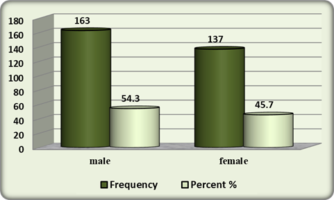
Figure (1) Distribution of the sample according to the incidence of Gender
Table (2): Distribution of the sample according to the incidence of age:
|
Age |
Frequency |
Percentage % |
|
20 to less than 30 years |
11 |
3.7 |
|
30 to less than 40 years |
47 |
15.7 |
|
40 to less than 50 years |
69 |
23 |
|
50 to less than 60 years |
53 |
17.7 |
|
60 to less than 70 years |
53 |
17.7 |
|
70 years and more |
67 |
22.3 |
|
total |
300 |
100 |
The data set out in the above table relating to the distribution of the sample individuals on the basis of age indicated that 3.7% of the sample were aged 20 to less than 30 years, 15.7% of the sample were aged 30 to less than 40 years, 23% of the sample ranged in age from 40 to less than 50 years, 17.7% of the sample were aged 50 to less than 60 years, 17.7% were aged from 60 to less than 70 years, and 22.3% of the sample ranged in age from 70 years and more.
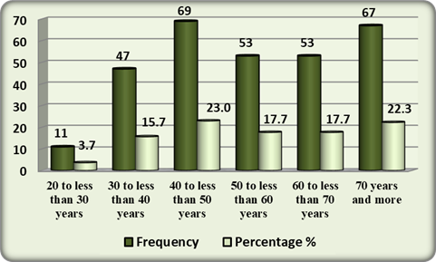
Figure (2) Distribution of the sample according to the incidence of Age
Table (3) : Distribution of the sample according to the incidence of Address:
|
Address |
Frequency |
Percentage % |
|
Al-Khoms |
174 |
58 |
|
Misallata |
126 |
42 |
|
Total |
300 |
100 |
As shown in Table 3, it was found that 58% of the respondents lived in the city of Al-Khoms, while 42% of them lived in the city of Misallata.
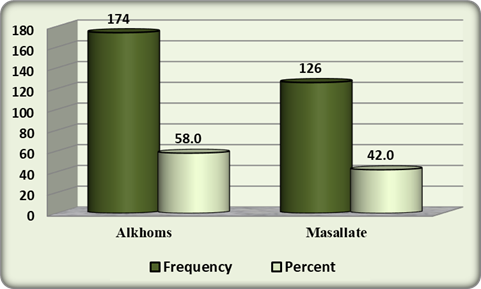
Figure (3) Distribution of the sample according to the incidence of address.
Table (4): Distribution of the sample according to the incidence of anemia:
|
anemia |
Frequency |
Percentage % |
|
Normal |
115 |
38.3 |
|
Abnormal |
288 |
61.7 |
|
Total |
300 |
100 |
From Table 4, it was found that 61.7% of the sample suffered from anemia, and it turned out that 38.3% of the sample did not have anemia, indicating that the prevalence of anemia was (61.7%).
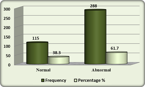
Figure (4) Distribution of the sample according to the incidence of anemia.
Table (5): Distribution of the sample according to the incidence of Diabetics:
|
Diabetics |
Frequency |
Percentage % |
|
Normal |
12 |
4 |
|
Abnormal |
288 |
96 |
|
Total |
300 |
100 |
From Table 5, it was found that (96%) of the sample suffer from diabetes, and it turns out that (4%) of the sample do not have diabetes.
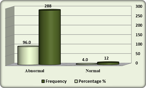
Figure (5) Distribution of the sample according to the incidence of diabetics.
Table 6: Results of Chi-square test to determine the differences in the incidence of anemia according to the gender:
|
Hb |
Total |
P-Value Sig. |
||||
|
Normal |
Abnormal |
|||||
|
Gender |
male |
No. |
71 |
92 |
163 |
0.028 |
|
Percentage |
43.6% |
56.4% |
100.0% |
|||
|
female |
No. |
44 |
93 |
137 |
||
|
Percentage |
32.1% |
67.9% |
100.0% |
|||
|
Total |
No. |
115 |
185 |
300 |
||
|
Percentage |
38.3% |
61.7% |
100% |
|||
|
Chi2 Calculated = 4.122 , df=1 , Chi2 Tabular 3.841 |
||||||
P-value <0.05 Significant , P-value <0.01 Highly significant , P-value > 0.05 non-Significant
The results showed that the P-value was 0.028, which was less than 0.05, indicating that there was a statistically significant difference in the incidence of anemia according to gender. The Chi² calculated value (4.122), which was greater than the tabular value (3.841), confirmed this relation. It was found that the percentage of females with anemia was 67.9%, which was higher than the percentage of males (56.4%).
Figure (6) the differences in the incidence of anemia according to the gender
Table 7: Results of Chi-square test to determine the differences in the incidence of anemia according to the age:
|
Hb |
Total |
P-Value Sig. |
|||||||
|
Normal |
Abnormal |
||||||||
|
age |
20 to less than 30 years |
No. |
7 |
4 |
11 |
0.072 |
|||
|
Percentage |
63.6% |
36.4% |
100.0% |
||||||
|
30 to less than 40 years |
No. |
24 |
23 |
47 |
|||||
|
Percentage |
51.1% |
48.9% |
100.0% |
||||||
|
40 to less than 50 years |
No. |
25 |
44 |
69 |
|||||
|
Percentage |
36.2% |
63.8% |
100.0% |
||||||
|
50 to less than 60 years |
No. |
21 |
32 |
53 |
|||||
|
Percentage |
39.6% |
60.4% |
100.0% |
||||||
|
60 to less than 70 years |
No. |
20 |
33 |
53 |
|||||
|
Percentage |
37.7% |
62.3% |
100.0% |
||||||
|
70years and more |
No. |
18 |
49 |
67 |
|||||
|
Percentage |
26.9% |
73.1% |
100.0% |
||||||
|
Total |
No. |
115 |
185 |
300 |
|||||
|
Percentage |
38.3% |
61.7% |
100.0% |
||||||
|
Chi2 Calculated = 10.103 , df =5 , Chi2 Tabular = 11.07 |
|||||||||
P-value <0.05 Significant , P-value <0.01 Highly significant , P-value > 0.05 non-Significant
The results showed that the P-value was 0.072, which was > 0.05, indicating that there were no statistically significant differences in the incidence of anemia according to age. The Chi² calculated value (10.103), which was less than the tabular value (11.07), confirms this result.
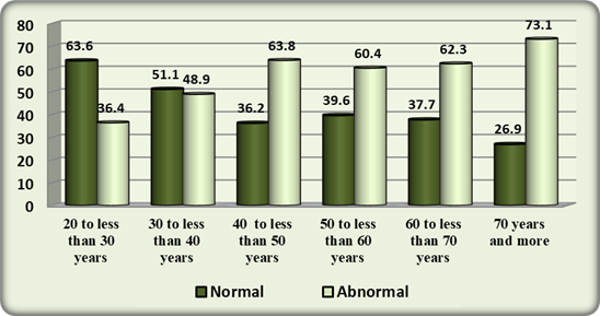
Figure (7) the differences in the incidence of anemia according to the age
Table 8: Results of Chi-square test to determine the differences in the incidence of anemia according to the place:
|
Hb |
Total |
P-Value Sig. |
|||||||
|
Normal |
Abnormal |
||||||||
|
Address |
Alkhoms |
No. |
74 |
100 |
174 |
0.051 |
|||
|
Percentage |
42.5% |
57.5% |
100.0% |
||||||
|
Misallata |
No. |
41 |
85 |
126 |
|||||
|
Percentage |
32.5% |
67.5% |
100.0% |
||||||
|
Total |
No. |
115 |
185 |
300 |
|||||
|
Percentage |
38.3% |
61.7% |
100.0% |
||||||
|
Chi2 Calculated = 3.085 , df=1 , Chi2 Tabular 3.841 |
|||||||||
P-value <0.05 Significant , P-value <0.01 Highly significant , P-value > 0.05 non-Significant
The results showed that the P-value was equal to 0.051, which was more than 0.05, and that there were no statistically significant differences in the incidence of anemia according to the place. The Chi² calculated value (3.085), which was less than the tabular value (3.841), confirms this result.
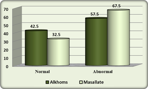
Figure (8) the differences in the incidence of anemia according to the place
Table 9: Results of Chi-square test to determine the differences in the incidence of anemia according to the diabetes:
|
Hb |
Total |
P-Value Sig. |
||||||||||
|
Normal |
Abnormal |
|||||||||||
|
HbAic |
Normal |
No. |
5 |
7 |
12 |
0.515 |
||||||
|
Percentage |
41.7% |
58.3% |
100.0% |
|||||||||
|
Abnormal |
No. |
110 |
178 |
288 |
||||||||
|
Percentage |
38.2% |
61.8% |
100.0% |
|||||||||
|
Total |
No. |
115 |
185 |
300 |
||||||||
|
Percentage |
38.3% |
61.7% |
100.0% |
|||||||||
|
Chi2 Calculated = 0.059 , df=1 , Chi2 Tabular 3.841 |
||||||||||||
P-value <0.05 Significant , P-value <0.01 Highly significant , P-value > 0.05 non-Significant
The results showed that the P-value was 0.515, which was greater than 0.05, and there were no statistically significant differences in the incidence of anemia according to diabetes. The Chi² calculated value (0.059), which was less than the tabular value (3.841), confirms this result. The researchers believe that this was due to the high incidence of diabetes, which included anemic and non-anemic patients.
Discussion
Diabetes is a major public health issue worldwide. The global burden of diabetes is increasing and is expected to be approximately 366 million by 2030 (12) . Anemia is common in diabetes and potentially contributes to the pathogenesis of diabetes complications (13). In our study, we included 300 patients with DM (163 men, 54.3%) and (137 women, 45.7%), with a mean age of 45 years. It was found that the prevalence of anemia in our study population was 61.7%, and the percentage of diabetics in the study area was 96%. In this research there were differences in the incidence of anemia according to gender, and it was found that the percentage of females with anemia was higher than the percentage of males.
The study concluded that there were no differences in the incidence of anemia according to the place of residence in Al-Khoms or Misallata and the incidence of diabetes. However, our prevalence estimate was higher than those reported in studies conducted elsewhere, including 14.6% in a tertiary center in Germany, 23.3% in the Austin and Repatriation Medical Center, Australia, and 15% and 23.5% in the diabetes care clinics of Teesside and Liverpool, UK (14). The prevalence of anemia in our study (61.7%) was relatively higher than that reported in the general adult population in Cameroon (34%) (15). The prevalence of anemia in patients with diabetes reported elsewhere is lower than our findings, depending on the level of development of the country, geographical altitude, and age of the study population: 46.5% in the Caribbean population, 17% in Ethiopia, and 12–23% in Caucasians(16).
Conclusion
The main objective of this study was to determine the prevalence of anemia in type 2 diabetic patients in Misallata and Alkhoms cities. In conclusion, the prevalence of anemia in the type 2 diabetic population in our study was high, including in patients with normal renal function. Anemia, characterized by a decline in RBC and Hb levels, is associated with diabetes, which, in turn, can itself result in anemia by virtue of multiple pathophysiological pathways, such as inflammation and decline in EPO. Female sex and advancing age are risk factors for the development of anemia in patients with diabetes. Similarly, the morphological type of anemia in diabetics may vary. The majority of patients had moderate anemia, and females suffered more from anemia than males. Our findings highlight the need to incorporate anemia screening into the routine assessment of diabetic complications, particularly for those with significant risk factors, to enable early detection and treatment of anemia and, hence, improve the overall care of patients with diabetes.
References
1- J. E. Shaw, R. A. Sicree, and P. Z. Zimmet, “Global estimates of the prevalence of diabetes for 2010 and 2030,” Diabetes Research and Clinical Practice, vol. 87, no. 1, pp. 4–14, 2010.
2- P. F. Pereira, R. D. C. G. Alfenas, and R. M. A. Ara´ujo, “Does breastfeeding influence the risk of developing diabetes mellitus in children? A review of current evidence,” Jornal de Pediatria, vol. 90, no. 1, pp. 7–15, 2014.
3-BrasilMinist´erio da Sa´ude, Diretrizes da Sociedade Brasileira de Diabetes 2013-2014, AC Farmacˆeutica, 2014.
4- A. Angelousi and E. Larger, “Anaemia, a common but often unrecognized risk in diabetic patients: a review, Diabetes & Metabolism, vol. 41, no. 1, pp. 18–27, 2015.
5- McGill JB, Bell DS Anemia and the role of erythropoietin in diabetes. J diabetes complications.2006; 20(4):262-72.
6- World Health Organization, Centers for Disease Control and Prevention. Assessing the iron status of populations. 2007. Available at: URL: http://www.who.int/nutrition/publications/micronutrients/ anaemia_iron_deficiency/9789241596107/en/
7- S. Fava, J. Azzopardi, S. Ellard, and A. T. Hattersley, “ACE gene polymorphism as a prognostic indicator in patients with type 2 diabetes and established renal disease,” Diabetes Care, vol. 24, no. 12, pp. 2115–2120, 2001.
8- WHO DIAMOND Project Group: Familial IDDM epidemiology: Standardization of data for the DIAMOND Project. Bull WHO 69:767-77, 2012.
9- WHO, Anaemia, World Health Organization, 2001.
10-WHO and IDF (2006):definition and diagnosis of diabetes mellitus and intermediate hyperglycemia: report of WHO/IDF consultation. WHO Document Production Services, Geneva, Switzerland.
11- World Health Organization, Centers for Disease Control and Prevention. Assessing the iron status of populations. 2007. Available at: URL: http://www.who.int/nutrition/publications/micronutrients/ anaemia_iron_deficiency/9789241596107/en/
12- Barbieri J, Fontela PC, Winkelmann ER, Zimmermann CEP, Sandri YP ,Viera EK et al. Anemia in Patients with Type 2 Diabetes Mellitus. Anemia 2015. pageshttp://dx.doi.org/10.1155/ 2015/354737.
13- Andrews M and Arredondo M. Ferritin levels and hepcidin mRNA expression in peripheral mononuclear cells from anemic type 2 diabetic patients. Biol Trace Elem Res.2012; 149(1):1-4.
14-M. C. Carvalho, E. C. E. Baracat, and V. C. Sgarbieri, “Anemia ferropriva e anemia de doenc¸a crˆonica: dist´urbios do metabolismo de ferro,” Revista Seguranc¸a Alimentar e Nutricional, vol. 13, no. 2, pp. 54–63, 2006.
15-M. Andrews and M. Arredondo, “Ferritin levels and hepcidin mRNA expression in peripheral mononuclear cells from anemic type 2 diabetic patients,” Biological Trace Element Research, vol. 149, no. 1, pp. 1–4, 2012.
16- D. K. Singh, P. Winocour, and K. Farrington, “Erythropoietic stress and anemia in diabetes mellitus,” Nature Reviews Endocrinology, vol. 5, no. 4, pp. 204–210, 2009.