

Comparison between Anti-VEGF (Bevacizumab) and Corticosteroid (Triamcinolone Acetonide) in the Treatment of Refractory Diabetic Macular Edema
مقارنة بين مضاد عامل نمو بطانة الأوعية الدموية (بيفاسيزوماب) والكورتيكوستيرويد (تريامسينولون أسيتونيد) في علاج الوذمة البقعية السكرية المقاومة للعلاج
Rawan Benayad1, Algharib Alshraa1, Rania Alkhoja1, Maison Daloub1, Juma Omrani1
¹ Ophthalmology Department, Libyan Board for Postgraduate Studies, Libya.
DOI: https://doi.org/10.53796/hnsj711/9
Arabic Scientific Research Identifier: https://arsri.org/10000/711/9
Volume (7) Issue (11). Pages: 191 - 201
Received at: 2026-10-05 | Accepted at: 2026-10-12 | Published at: 2026-11-01
Abstract: Objective: To compare the effectiveness and ocular safety of intravitreal bevacizumab and triamcinolone acetonide in refractory diabetic macular edema. Methods: This retrospective comparative study at Tripoli Eye Hospital, Libya, included 50 patients with an insufficient response to three previous bevacizumab injections. Twenty-five received three monthly bevacizumab injections (1.25 mg/0.05 mL), and 25 received one triamcinolone injection (4 mg/0.1 mL). Central macular thickness (CMT), best-corrected visual acuity (BCVA), and ocular complications were assessed over six months. Results: At three months, mean CMT decreased from 418 to 315 µm with bevacizumab and from 420 to 255 µm with triamcinolone; mean BCVA increased from 0.15 to 0.25 and 0.50, respectively. Between-group differences favored triamcinolone at one and three months. At six months, CMT was 285 versus 275 µm (p = 0.47), and BCVA was 0.40 in both groups (p = 1.00). Intraocular pressure elevation and cataract occurred in 32% and 24% with triamcinolone versus 8% and 4% with bevacizumab. Conclusion: Triamcinolone provided greater early improvement, but no significant between-group differences remained at six months. Its higher frequency of ocular complications warrants careful monitoring.
Keywords: Diabetic macular edema; Bevacizumab; Triamcinolone; Macular thickness; Visual acuity.
المستخلص: الهدف: مقارنة فعالية وسلامة حقن البيفاسيزوماب والتريامسينولون أسيتونيد داخل الجسم الزجاجي لعلاج الوذمة البقعية السكرية المقاومة للعلاج. المنهجية: شملت دراسة مقارنة استعادية بمستشفى طرابلس للعيون، ليبيا، ٥٠ مريضًا لم يستجيبوا استجابة كافية لثلاث حقن سابقة من البيفاسيزوماب. تلقى ٢٥ مريضًا ثلاث حقن شهرية من البيفاسيزوماب (١٫٢٥ ملغ/٠٫٠٥ مل)، وتلقى ٢٥ مريضًا حقنة واحدة من التريامسينولون (٤ ملغ/٠٫١ مل). قُيّمت سماكة البقعة المركزية وأفضل حدة إبصار مصححة والمضاعفات خلال ستة أشهر. النتائج: بعد ثلاثة أشهر، انخفض متوسط السماكة من ٤١٨ إلى ٣١٥ ميكرومتر مع البيفاسيزوماب، ومن ٤٢٠ إلى ٢٥٥ مع التريامسينولون، وتحسنت حدة الإبصار من ٠٫١٥ إلى ٠٫٢٥ و٠٫٥٠، على التوالي. رجحت الفروق كفة التريامسينولون بعد شهر وثلاثة أشهر. بعد ستة أشهر، بلغت السماكة ٢٨٥ مقابل ٢٧٥ ميكرومتر (p = 0.47)، وحدّة الإبصار ٠٫٤٠ بالمجموعتين (p = 1.00). بلغ ارتفاع ضغط العين والساد ٣٢٪ و٢٤٪ مع التريامسينولون مقابل ٨٪ و٤٪ مع البيفاسيزوماب. الاستنتاج: حقق التريامسينولون تحسنًا مبكرًا أكبر، دون فروق دالة بين المجموعتين بعد ستة أشهر، وتستدعي زيادة مضاعفاته مراقبة دقيقة.
الكلمات المفتاحية: الوذمة البقعية السكرية؛ بيفاسيزوماب؛ تريامسينولون؛ سماكة البقعة؛ حدة الإبصار.
INTRODUCTION:
Diabetes mellitus is a chronic disease that affects 415 million people worldwide, despite treatment advances diabetic eye disease remains a major cause of vision loss worldwide.
Diabetic macular edema (DME) is a retinal manifestation of diabetes characterized by accumulation of fluid within the macula as a consequence of disruption of the blood-retinal barrier. It is a major cause of visual impairment in people with diabetic retinopathy.(1) (2)
Chronic hyperglycemia induces biochemical and cellular abnormalities, including pericyte loss, endothelial dysfunction, oxidative stress, retinal ischemia, and disruption of endothelial tight junctions. These changes increase vascular permeability and permit plasma fluid and proteins to enter the retinal extracellular space, producing retinal thickening and cystoid changes on optical coherence tomography (OCT).(3,4) (2)
The pathogenesis of DME is multifactorial rather than being driven by a single mediator. Vascular endothelial growth factor (VEGF) has a central role because hypoxia, ischemia, and inflammatory signaling increase VEGF expression, which promotes vascular leakage and breakdown of the blood-retinal barrier.(2,5)
At the same time, DME has an important inflammatory component. Cytokines and chemokines, leukocyte activation, and dysfunction of the retinal neurovascular unit can sustain vascular leakage and edema. VEGF and inflammation interact with each other, which helps explain why suppression of VEGF alone does not completely resolve edema in every patient. (2)
Treatment of center-involving DME has evolved from focal/grid laser photocoagulation to intravitreal pharmacologic therapy. Anti-VEGF treatment is currently the principal first-line therapy for center-involving DME because randomized trials have demonstrated superior visual outcomes compared with laser alone.(1) (5)
Common anti-VEGF agents used in DME include bevacizumab, ranibizumab, and aflibercept.
Bevacizumab (Avastin) is a full-size humanized recombinant monoclonal immunoglobulin G antibody that inactivates all VEGF isoforms .(7)
It is widely used as a first-line anti-VEGF option in clinical practice because it has demonstrated efficacy and has a substantially lower treatment cost than several licensed alternatives. In the DRCR.net Protocol T trial, bevacizumab, ranibizumab, and aflibercept all improved visual acuity; the relative advantage of aflibercept was mainly seen in eyes with worse baseline vision. Thus, bevacizumab remains a practical first-line option in many resource-limited settings when cost and access are important considerations.(8) (6)
Not all eyes achieve complete resolution of DME with anti-VEGF therapy. Persistent or refractory DME is not uniformly defined, but many studies describe it as edema that remains despite an adequate course of anti-VEGF treatment.(5)
Expert consensus definition based on diabetic retinopathy clinical research network (DRCR.met) was persistent center-involving DME after three consecutive monthly intravitreal anti-VEGF injections with a visual acuity improvement of ≤ 5 ETDRS letters and ≤ 10% reduction in central foveal thickness from baseline as assessed by OCT .(9)
Persistent edema may be related to chronic inflammation inflammatory mediators and cytokines including interleukin-6, tumor necrosis and monocyte chemoattractant protein have been implicated in disruption of blood retinal barrier, also structural retinal damage,( disorganization of the retinal inner layer Drill, ellipsoid zone disruption photoreceptor damage, chronic intraretinal cysts, disruption of the outer retinal layers) inadequate treatment exposure, or mechanisms that are not exclusively VEGF-dependent.(4)
The number of anti-VEGF injections required before judging response is also variable. A three-injection monthly loading phase is commonly used, but some treatment
strategies extend the loading phase to six monthly injections when edema persists.(10)
Evidence from Protocol T post-hoc analyses showed that some eyes that remained edematous after the initial 12-week period continued to improve through week 24, supporting the concept that DME may require a longer period of anti-VEGF therapy.(11)
When edema persists despite adequate anti-VEGF therapy, switching to another anti-VEGF agent, using a corticosteroid, combination therapy, laser in selected situations.
Corticosteroids are particularly relevant because they target inflammatory pathways as well as vascular permeability. Triamcinolone acetonide (Kenacort) is a potent
Corticosteroid that suppresses inflammatory mediators and reduces vascular permeability, thereby decreasing retinal fluid and thickness. This mechanism is clinically relevant because inflammation is an important component of DME, especially in chronic or treatment-resistant disease.
However, intravitreal triamcinolone has important limitations. Its effect is temporary, and repeated exposure may cause intraocular pressure elevation and cataract progression.(12)
These adverse effects must be balanced against the potential for a rapid anatomical response in selected refractory eyes. Comparative studies, including the IBEME study by Paccola et al. and the randomized trial by Rodrigues et al., have evaluated bevacizumab and triamcinolone in persistent or refractory DME and provide a clinical basis for comparing their anatomical, functional, and safety profiles.
2. LITERATURE REVIEW
Paccola et al. (IBEME study) (2008) conducted a randomized study in 28 eyes comparing a single intravitreal injection of triamcinolone acetonide( 4mg) with bevacizumab( 1.5mg) in refractory diffuse DME. The study reported significantly greater reduction in CMT with triamcinolone at weeks 4, 8, 12, 24. However, the significant improvement in BCVA with triamcinolone was observed only at weeks 8, 12, with no significant difference between the groups at week 24.(13)
Azad et al. (2012) conducted a prospective randomized study in 60 eyes compared intravitreal bevacizumab, intravitreal triamcinolone acetonide, and macular grid augmentation in refractory diffuse DME. At 6 months both bevacizumab and triamcinolone acetonide groups showed significant improvement in visual acuity and central macular thickness which is nearly the same in both groups, but the laser group showed only marginal improvement in visual acuity. VT associated with side effects such as IOP rise and cataract formation.(14)
Shimura et al.(2008) A prospective comparative study in 30 eyes compared bevacizumab and triamcinolone acetonide in persistent diffuse DME and reported IVT showed better results in reducing DME and improvement of VA than of bevacizumab in early follow-up suggesting that the pathogenesis of DME is not only attributed to VEGF-dependency but also attributed to other inflammatory mechanism suppressed by corticosteroid. And both groups showed the recurrence of macular edema at 24 week but triamcinolone acetonide kept better results than bevacizumab. (15)
Rodrigues et al (2020) conducted a randomized clinical trial in 100 eyes comparing continued treatment with prn-IVB with a switch to Prn-IVT in eyes with persistent diabetic macular edema after bevacizumab treatment. At week 48, there was no statistically significant difference between the two groups in central macular thickness or BCVA However triamcinolone treatment was associated with a higher risk of intraocular pressure elevation .(16)
Mohanty et al. (2019) conducted a prospective randomized clinical trial comparing intravitreal bevacizumab with intravitreal triamcinolone acetoinde in 52 eyes with recalcitrant diabetic macular edema, both treatment groups produced comparable visual improvement at 24 week with triamcinolone was associated with higher risk of intraocular pressure elevation. (17)
3. AIM OF THE STUDY
- To compare the anatomical and functional outcomes between the two treatment groups.
- To assess ocular complications of intravitreal bevacizumab versus intravitreal triamcinolone acetonide in patients with refractory diabetic macular edema.
4. OBJECTIVES
- To compare changes in central macular thickness (CMT) between the two treatment groups.
- To compare changes in best-corrected visual acuity (BCVA) between the two treatment groups.
- To assess clinically relevant ocular adverse effects, particularly intraocular pressure elevation and cataract progression.
5. MATERIALS AND METHODS:
5.1 Study Design
A retrospective comparative study.
5.2 Study Setting
Tripoli Eye Hospital, Libya.
5.3 Study Population
Fifty adult patients with refractory diabetic macular edema were included: 25 patients treated with Avastin (bevacizumab) and 25 patients treated with Kenacort (triamcinolone acetonide).
5.4 Inclusion Criteria
- Age ≥ 18 years.
- Type 1 or type 2 diabetes mellitus.
- Diabetic macular edema confirmed clinically and by OCT.
- Baseline CMT ≥ 300 µm.
- Persistent/refractory edema despite previous at least 3 injections of anti-VEGF therapy.
- Adequate baseline and follow-up data.
5.5 Exclusion Criteria
- Other significant ocular disease affecting visual outcome, such as age-related macular degeneration or retinal vein occlusion.
- Previous vitrectomy.
- Active ocular infection or intraocular inflammation.
- Significant media opacity affecting OCT or visual assessment.
- Advanced or uncontrolled glaucoma.
- Incomplete clinical or follow-up data.
5.6 Data Collection
- Age and sex.
- Duration of diabetes and HbA1c.
- Previous anti-VEGF treatment.
- BCVA and CMT.
- IOP.
- Cataract.
- Endophthalmitis.
- Retinal detachment.
5.7 Treatment Protocol
Avastin group: intravitreal bevacizumab 1.25 mg/0.05 mL, administered as three injections at one-month intervals. Kenacort group: intravitreal triamcinolone acetonide 4 mg/0.1 mL, administered as a single intravitreal injection.
5.8 Outcome Measures
The primary outcomes were changes in CMT and BCVA. IOP elevation, cataract progression, endophthalmitis, and retinal detachment were assessed as safety outcomes.
5.9 Statistical analysis:
Data were analyzed using SPSS.
6. RESULTS:
Table 1. Baseline demographic and clinical characteristics:
| Variable | Avastin (n=25) | Kenacort (n=25) |
| Mean age (years) | 60.2 ± 8.1 | 60.7 ± 7.8 |
| Male | 12 (48%) | 15 (60%) |
| Female | 13 (52%) | 10 (40%) |
| Mean Duration of diabetes (years) | 12.1 ± 5.0 | 12.7 ± 5.1 |
| Mean HbA1c (%) | 7.5 ± 0.9 | 7.6 ± 0.8 |
| Previous anti-VEGF injections | 3 | 3 |
| Mean Baseline IOP (mmHg) | 15.7 ± 2.4 | 15.9 ± 2.5 |
The baseline demographic and clinical characteristics were comparable between the two treatment groups.
6.2 Best corrected visual acuity
Table 2. Mean central macular thickness during follow-up
| Follow-up | Avastin CMT (µm) | Kenacort CMT (µm) | P value |
| Baseline | 418 ± 57 | 420 ± 49 | 0.89 |
| 1 month | 345 ± 48 | 305 ± 41 | 0.003 |
| 3 months | 315 ± 40 | 255 ± 39 | < 0.001 |
| 6 months | 285 ± 45 | 275 ± 50 | 0.47 |
Mean CMT decreased in both groups. In the Avastin group, CMT decreased from
418 ± 57 µm at baseline to 345 ± 48 µm at 1 month and 315 ± 40 µm at 3 months, reaching 285 ± 45 µm at 6 months. In the Kenacort group, CMT decreased from 420 ± 49 µm at baseline to 305 ± 41 µm at 1 month and 255 ± 39 µm at 3 months, followed by an increase to 275 ± 50 µm at 6 months. The between-group difference was statistically significant at 1 month (p = 0.003) and 3 months (p < 0.001), with no significant difference between the groups at 6 month.
Table 3. Mean best-corrected visual acuity during follow-up
| Follow-up | Avastin BCVA | Kenacort BCVA | P value |
| Baseline | 0.15 ± 0.025 | 0.15 ± 0.05 | 0.9 |
| 1 month | 0.20 ± 0.10 | 0.30 ± 0.05 | < 0.001 |
| 3 months | 0.25 ± 0.10 | 0.50 ± 0.025 | < 0.001 |
| 6 months | 0.40 ± 0.025 | 0.4 ± 0.05 | 1.00 |
6.3 Safety Outcomes
Table 4. Ocular complications during follow-up
| Outcome | Avastin (n=25) | Kenacort (n=25) |
| IOP elevation | 2 (8%) | 8 (32%) |
| Cataract | 1 (4%) | 6 (24%) |
| Endophthalmitis | 0 | 0 |
| Retinal detachment | 0 | 0 |
BCVA improved in both groups during follow-up. In the Avastin group, mean BCVA increased from 0.15 ± 0.025 at baseline to 0.20 ± 0.10 at 1 month, 0.25 ± 0.10 at 3 months, and 0.40 ± 0.025 at 6 months. In the Kenacort group, BCVA increased from 0.15 ± 0.05 at baseline to 0.30 ± 0.05 at 1 month and 0.50 ± 0.025 at 3 months, followed by a decrease to 0.4 ± 0.05 at 6 months. The between-group differences were statistically significant at 1 month (p < 0.001), 3 months (p < 0.001). With no significant difference between the groups at 6 month.
IOP elevation and cataract were more frequent in the Kenacort group (32% and 24%, respectively) than in the Avastin (8%, 4% respectively), no cases of endophthalmitis or retinal detachment were recorded.
6.4 figures
Figures 1 and 2. Changes in central macular thickness (CMT) and best-corrected visual acuity (BCVA) over 6 months.
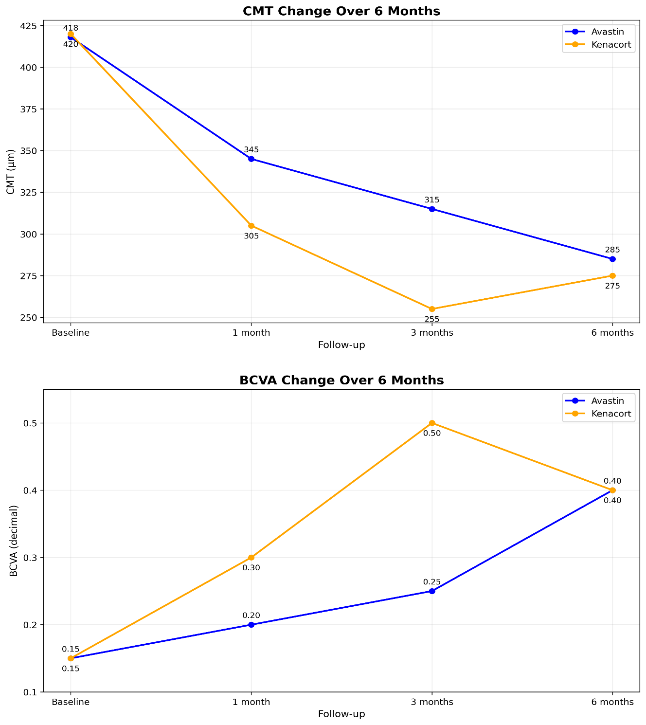
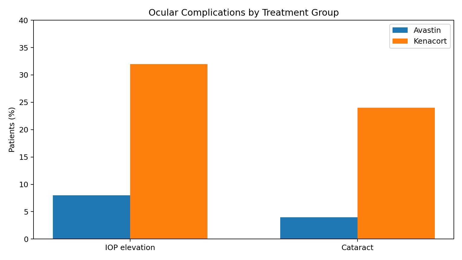
Figure 3. Frequency of intraocular pressure (IOP) elevation and cataract by treatment group.
7. DISCUSSION:
The preset compare the effects of intravitreal bevacizumab (avastin) and intravitreal triamcinolone acetonide (kenacort) in patients with refractory diabetic macular edema. In our study kenacort produced a significantly greater improvement in visual acuity and significantly improvement in reduction of central macular thickness (CMT) at the first and third months compared with avastin. At 6 months, however, the difference between the two groups was no longer statistically significant.
The anatomical response in the kenacort group showed an important pattern during follow-up. CMT decreased markedly during the first three months, followed by rebound increase at six months, despite this rebound the CMT at six months remained numerically lower in the kenacort group than in the avastin group this may indicate that the early anatomical effect of kenacort was stronger, but its effect decreased with time after a single injection.
Our findings are in agreement with paccola et al. who studied refractory DME and found a significantly greater reduction in CMT with triamcinolone at weeks 8 and 12, while the difference between the two groups was no longer significant at week 24.
The results of Azad et al. also support the anatomical findings of our study .AT 6 months both treatments significantly improved visual acuity and reduced CMT, with greater numerical reduction in CMT in the triamcinolone group compared with bevacizumab group.
Shimura et al. similarly reported a stronger effect of triamcinolone on macular edema and visual acuity , both treatment initially reduced the edema , but recurrence occurred with time while the triamcinolone treated eyes maintained a better anatomical and visual result during follow-up.
In contrast Mohanty et al. found that bevacizumab and triamcinolone were similarly effective in improving visual acuity and reduced CMT in refractory DME their findings agree with our six month results, when the difference between the two groups was no longer statistically significant.
Rodrigues al. also reported no significant difference between continued bevacizumab and switching to triamcinolone in patients with refractory DME after previous bevacizumab treatment. At 48 weeks, there was no significant difference in either CMT or visual acuity between the two groups.
The stronger early advantage of the kenacort observed in our study may be related to its broader anti-inflammatory effect in addition to reducing vascular permeability, in refractory DME inflammatory mechanisms may contribute to persist retinal thickening despite anti-VEGF treatment. The rebound in CMT at six months suggests that the effect of a single triamcinolone injection may decrease with time.
Overall, our results suggest that kenacort produced a stronger early anatomical and functional response than avastin in refractory DME, particularly during the first three months. By the six months, this difference was no longer statistically significant, although CMT remained numerically lower in kenacort group, these findings are generally consistent with previous studies showing a stronger early response to triamcinolone followed by a reduction of the treatment difference during longer follow-up. Also the use of kenacort should be considered carefully because of its potential ocular complication, in our study elevation of intraocular pressure and cataract progression was more frequent in kenacort group, these findings are consistent with the known ocular side effect of intravitreal corticosteroids, therefore kenacort may be in selected cases in refractory diabetic macular edema
Limitation:
This study has some limitations, including retrospective design relatively small sample size and short follow-up period.
8. CONCLUSION
Triamcinolone acetonide demonstrated greater early improvement in both visual acuity and central macular thickness compared with bevacizumab .However, a rebound in central macular thickness was observed at the final follow-up, resulting in comparable visual and anatomical outcomes between the two groups .although CMT remained numerically lower in the triamcinolone group at 6 months but the difference was not statistically significant.
Recommendation:
Intravitreal triamcinolone acetonide may be considered as an alternative treatment option in selected patients with refractory diabetic macular edema, particularly in cases with persistent edema despite previous anti-VEGF therapy. However, its use should be accompanied by careful monitoring of intraocular pressure and cataract development. Further prospective studies with larger sample size and longer follow-up are recommended to confirm these findings and to better define the patients who may benefit most from corticosteroid therapy.
9. REFERENCES
- Barham R, El Rami H, Sun JK, Silva PS. Evidence-Based Treatment of Diabetic Macular Edema. Semin Ophthalmol. 2017;32(1):56–66. doi:10.1080/08820538.2016.1228388 PubMed PMID: 28060586.
- Noma H, Yasuda K, Shimura M. Involvement of Cytokines in the Pathogenesis of Diabetic Macular Edema. Int J Mol Sci. 2021 Mar 26;22(7). doi:10.3390/ijms22073427 PubMed PMID: 33810434; PubMed Central PMCID: PMC8036935.
- Das A, McGuire PG, Rangasamy S. Diabetic Macular Edema: Pathophysiology and Novel Therapeutic Targets. Ophthalmology. 2015 Jul;122(7):1375–94. doi:10.1016/j.ophtha.2015.03.024 PubMed PMID: 25935789.
- Rübsam A, Parikh S, Fort PE. Role of Inflammation in Diabetic Retinopathy. Int J Mol Sci. 2018 Mar 22;19(4). doi:10.3390/ijms19040942 PubMed PMID: 29565290; PubMed Central PMCID: PMC5979417.
- Shah SU, Maturi RK. Therapeutic Options in Refractory Diabetic Macular Oedema. Drugs. 2017 Apr;77(5):481–92. doi:10.1007/s40265-017-0704-6 PubMed PMID: 28197794.
- Wells JA, Glassman AR, Ayala AR, Jampol LM, Bressler NM, Bressler SB, et al. Aflibercept, Bevacizumab, or Ranibizumab for Diabetic Macular Edema: Two-Year Results from a Comparative Effectiveness Randomized Clinical Trial. Ophthalmology. 2016 Jun;123(6):1351–9. doi:10.1016/j.ophtha.2016.02.022 PubMed PMID: 26935357; PubMed Central PMCID: PMC4877252.
- Stefanini FR, Arevalo JF, Maia M. Bevacizumab for the management of diabetic macular edema. World J Diabetes. 2013 Apr 15;4(2):19–26. doi:10.4239/wjd.v4.i2.19 PubMed PMID: 23593532; PubMed Central PMCID: PMC3627413.
- Ross EL, Hutton DW, Stein JD, Bressler NM, Jampol LM, Glassman AR. Cost-effectiveness of Aflibercept, Bevacizumab, and Ranibizumab for Diabetic Macular Edema Treatment: Analysis From the Diabetic Retinopathy Clinical Research Network Comparative Effectiveness Trial. JAMA Ophthalmol. 2016 Aug 1;134(8):888–96. doi:10.1001/jamaophthalmol.2016.1669 PubMed PMID: 27280850; PubMed Central PMCID: PMC6648661.
- Sorour OA, Levine ES, Baumal CR, Elnahry AG, Braun P, Girgis J, et al. Persistent diabetic macular edema: Definition, incidence, biomarkers, and treatment methods. Surv Ophthalmol. 2023 Apr;68(2):147–74. doi:10.1016/j.survophthal.2022.11.008 PubMed PMID: 36436614.
- Rehmani A, Banaee T, Alwan S, Urias E, Lyons L, El-Annan J. Pro re nata Treatment of Diabetic Macular Edema with Cycles of Three Injections of Anti-vascular Endothelial Growth Factor Injections. Middle East Afr J Ophthalmol. 2023 Sep;30(3):141–8. doi:10.4103/meajo.meajo_17_22 PubMed PMID: 39444996; PubMed Central PMCID: PMC11495293.
- Udaondo P, Adan A, Arias-Barquet L, Ascaso FJ, Cabrera-López F, Castro-Navarro V, et al. Challenges in Diabetic Macular Edema Management: An Expert Consensus Report. Clin Ophthalmol Auckl NZ. 2021;15:3183–95. doi:10.2147/OPTH.S320948 PubMed PMID: 34349495; PubMed Central PMCID: PMC8327476.
- Whitcup SM, Cidlowski JA, Csaky KG, Ambati J. Pharmacology of Corticosteroids for Diabetic Macular Edema. Invest Ophthalmol Vis Sci. 2018 Jan 1;59(1):1–12. doi:10.1167/iovs.17-22259 PubMed PMID: 29297055; PubMed Central PMCID: PMC5961100.
- Paccola L, Costa RA, Folgosa MS, Barbosa JC, Scott IU, Jorge R. Intravitreal triamcinolone versus bevacizumab for treatment of refractory diabetic macular oedema (IBEME study). Br J Ophthalmol. 2008 Jan;92(1):76–80. doi:10.1136/bjo.2007.129122 PubMed PMID: 17965109.
- Azad R, Sain S, Sharma YR, Mahajan D. Comparison of intravitreal bevacizumab, intravitreal triamcinolone acetonide, and macular grid augmentation in refractory diffuse diabetic macular edema: A prospective, randomized study. Oman J Ophthalmol. 2012 Sep;5(3):166–70. doi:10.4103/0974-620X.106100 PubMed PMID: 23439853; PubMed Central PMCID: PMC3574512.
- Shimura M, Nakazawa T, Yasuda K, Shiono T, Iida T, Sakamoto T, et al. Comparative therapy evaluation of intravitreal bevacizumab and triamcinolone acetonide on persistent diffuse diabetic macular edema. Am J Ophthalmol. 2008 May;145(5):854–61. doi:10.1016/j.ajo.2007.12.031 PubMed PMID: 18328456.
- Rodrigues MW, Cardillo JA, Messias A, Siqueira RC, Scott IU, Jorge R. Bevacizumab versus triamcinolone for persistent diabetic macular edema: a randomized clinical trial. Graefes Arch Clin Exp Ophthalmol Albrecht Von Graefes Arch Klin Exp Ophthalmol. 2020 Mar;258(3):479–90. doi:10.1007/s00417-019-04564-z PubMed PMID: 31873786.
- muhanty. comprative study between intravitreal bevacizumab and intravitreal triamcinolon in recalcitrant.
 Arabic
 English